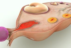
Placenta accreta is the attachment of the placenta to the innermost layers of the uterus, uterine muscle or even outside the uterus in organs such as the bladder or intestine, for example, and is a serious complication during pregnancy, as it can make it difficult for the placenta to exit during childbirth. and cause postpartum hemorrhage.
Placenta accreta is typically identified on prenatal ultrasounds and is most commonly caused due to previous cesarean sections. However, other factors can contribute to its development, such as advanced maternal age, uterine curettage, or placenta previa, for example.
It is important that placenta accreta, also known as placental accreta, is diagnosed during prenatal examinations so that a cesarean section can be scheduled followed by hysterectomy, which is normally the recommended treatment, and thus complications for the mother and for the baby.

Placenta accreta symptoms
Normally, women do not feel any symptoms of changes in the placenta, which is why it is important that women carry out prenatal care correctly so that this change can be identified.
Although signs and symptoms are not frequent in these cases, some women may experience discrete vaginal bleeding, without pain and for no apparent reason during pregnancy, and it is recommended that they go to the gynecologist/obstetrician to identify the cause of the bleeding and begin treatment. .
How the diagnosis is made
The diagnosis of placenta accreta is made by the obstetrician through imaging tests, such as prenatal ultrasound, allowing the doctor to visualize abnormalities in the placenta, such as enlargement of blood vessels, as well as thinning of the myometrium and invasion of the placenta into the bladder, for example .
These tests can be carried out prenatally and early diagnosis of placental accreta reduces the risk of complications for women. Find out about other exams carried out during prenatal care.
Ultrasonography is normally indicated for women considered to be at high risk and is a very safe technique for both mother and baby.
In some cases, the doctor may request an MRI exam, which is controversial. However, it may be indicated when the ultrasound result is considered doubtful or inconclusive.
Types of placenta accreta
Placental accreta can be classified according to the depth of implantation of the placenta into the uterus into:
- Simple placenta accreta: it is the most common type, in which the placenta invades the decidua, which is the inner layer of the uterus;
- Placenta increscendo: in this type, the placenta completely penetrates the myometrium, which is the uterine muscle;
- Passed the cake: is the least common type, in which the placenta passes through the wall of the uterus, and can grow through the wall of the uterus and reach nearby organs, such as the bladder or intestine.
The type of placenta accreta is identified by the obstetrician through diagnostic tests.
Possible causes
Placental accreta is more common to be caused by previous cesarean surgeries, as loss of the decidua can occur in the cesarean scar, which is the part of the uterine mucosa where the placenta normally implants.
Some factors that may increase the risk of developing placental accreta are:
- Pregnancy after age 35;
- Multiple pregnancies;
- Fibroids;
- Surgeries to remove fibroids;
- Having previously performed curettage;
- Endometrial ablation anteriorly;
- Radiotherapy in the pelvic region.
Additionally, women who have placenta previa or a history of placenta previa in previous pregnancies also have an increased risk of developing placenta accreta. Understand what placenta previa is and how it is treated.
Possible risks
The risks of placenta accreta are related to when placenta accreta is identified. The sooner the diagnosis is made, the lower the risk of postpartum hemorrhage, complications during childbirth, premature birth and the need for an emergency cesarean section.
Furthermore, there may be infection, problems related to coagulation, bladder rupture, loss of fertility and, if not identified and treated correctly, it can lead to death.
For the baby, the risks are premature birth, or lack of oxygenation due to maternal hemorrhage.
How the treatment is carried out
The treatment of placental accreta is carried out by the obstetrician through cesarean section together with hysterectomy, which is the medical procedure in which the uterus is removed, normally between the 34th and the end of the 35th week of pregnancy, to optimize the baby’s maturity and reduce the risk of bleeding in women.
In cases where a woman experiences severe bleeding, the doctor may recommend a blood transfusion.
In some cases, conservative treatment may be indicated to preserve the woman’s fertility, with only a cesarean section and removal of the placenta, in addition to monitoring the woman after birth to monitor possible bleeding or complications.

Sign up for our newsletter and stay up to date with exclusive news
that can transform your routine!
Warning: Undefined array key "title" in /home/storelat/public_html/wp-content/plugins/link-whisper-premium/templates/frontend/related-posts.php on line 12
Warning: Undefined array key "title_tag" in /home/storelat/public_html/wp-content/plugins/link-whisper-premium/templates/frontend/related-posts.php on line 13