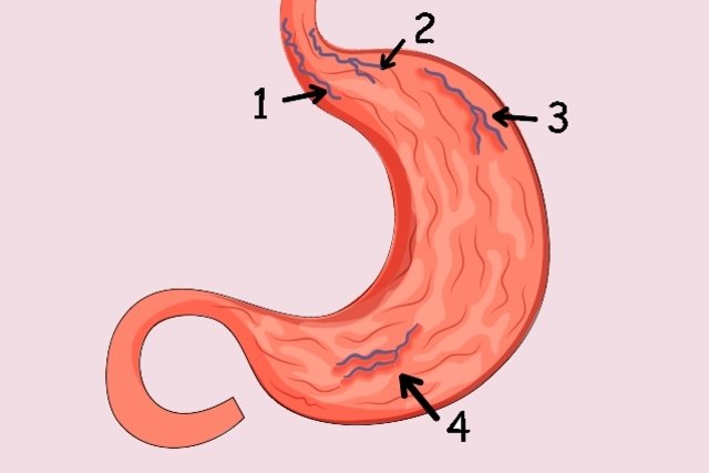
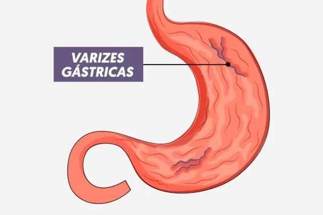
Varicose veins in the stomach, or gastric varices, are dilated and tortuous blood vessels that form in the wall of this organ, and can be serious, as as they become larger, they are at risk of rupturing and causing serious bleeding.
These varicose veins can form in the stomach due to increased blood flow resistance in the portal vein, which can be caused by chronic hepatitis, cirrhosis of the liver, schistosomiasis or thrombosis in the portal vein, for example.
Treatment for varicose veins in the stomach should be guided by a gastroenterologist or general practitioner and aims to prevent the appearance of new varicose veins, avoid stopping bleeding, which can be done with beta-blocker medications or surgical procedures, such as sclerotherapy, cyanoacrylate or elastic connectors, for example.
Main symptoms
The main symptoms of varicose veins in the stomach are:
- Dark, strong-smelling stools;
- Vomiting with blood;
- Paleness or yellowish skin;,
- Dizziness;
- Abdominal swelling;
- Weakness.
Symptoms of varicose veins in the stomach normally appear when the varicose veins rupture and, therefore, as soon as any sign or symptoms are noticed, the general practitioner or gastroenterologist is consulted so that tests can be carried out to confirm the diagnosis, identify the severity and initiate the most appropriate treatment.
How the diagnosis is made
The diagnosis of varicose veins in the stomach is made by a gastroenterologist or general practitioner by evaluating the signs and symptoms presented by the person and performing imaging tests, such as digestive endoscopy, Doppler ultrasound and tomography.
Make an appointment with your nearest gastroenterologist to assess your risk of stomach varicose veins:
Taking care of your health has never been easier!
After carrying out the tests, the doctor can classify the varicose veins according to their location and assess the size and risk of bleeding, which is essential to recommend treatment.
Gastric varices are considered small when they measure less than 3 mm in diameter, medium when they measure between 3 and 5 mm or large when they measure more than 5 mm in diameter. The larger the size of the varicose veins, the greater the risk of bleeding.
Causes of gastric varicose veins
The main causes of varicose veins in the stomach are:
- Chronic hepatitis;
- Hepatical cirrhosis;
- Schistosomiasis;
- Thrombosis of the Portal vein or splenic vein;
- Budd-Chiari syndrome;
- Malformations in the Portal vein or inferior vena cava.
Varicose veins in the stomach can also be caused by a heart disease called constrictive pericarditis, in which fibrous tissue develops around the heart, making it difficult to function.
How the treatment is carried out
The treatment of varicose veins in the stomach must be carried out according to the doctor’s instructions, and may vary depending on the location, size and risk of bleeding. If it is small or does not pose a risk of bleeding, regular monitoring is only recommended.
However, in some cases, treatment to prevent bleeding may be recommended, especially if they measure more than 10 mm in diameter or there is a serious risk of bleeding, which can be done with beta-blocker medications, which decrease the strength of the blood flow. , such as Propranolol, or the application of Cyanoacrylate, a type of glue that eliminates the vessel.
When gastric varices bleed, treatment may include endoscopy to perform sclerotherapy, injection of Cyanoacrylate or placement of elastic bandages, clips or springs, for example.

Bibliography
- Federle, Michael P. Imaging Diagnosis: Gastrointestinal. 1 ed. Rio de Janeiro: Elsevier, 2018. p. 114-117.

Sign up for our newsletter and stay up to date with exclusive news
that can transform your routine!
Warning: Undefined array key "title" in /home/storelat/public_html/wp-content/plugins/link-whisper-premium/templates/frontend/related-posts.php on line 12
Warning: Undefined array key "title_tag" in /home/storelat/public_html/wp-content/plugins/link-whisper-premium/templates/frontend/related-posts.php on line 13