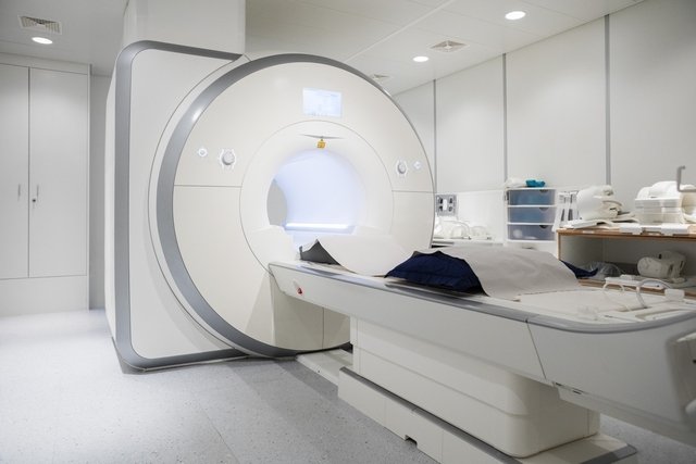
Magnetic resonance imaging (MRI) is a diagnostic exam that uses a high-intensity magnetic field and radio waves, allowing the internal structures of organs to be visualized with great definition, being able to identify various health problems, such as aneurysms, tumors, changes in joints or other injuries to internal organs.
This exam, also known as nuclear magnetic resonance (NMR), is requested by the doctor in cases of suspected changes in the brain, spinal cord, heart, bones, joints or other organs such as kidneys, liver or gallbladder, and in some cases it may be Gadolinium contrast was used to obtain higher quality images.
Magnetic resonance imaging can be carried out free of charge by the SUS, as long as it is medically indicated, but it is also carried out in hospitals or private examination clinics, and the results must be analyzed by the doctor who requested the examination.
What is it for
Magnetic resonance imaging is used to evaluate, identify, diagnose and monitor the treatment of various health conditions, such as:
- Neurodegenerative diseases such as Alzheimer’s or multiple sclerosis;
- Cerebrovascular diseases, such as brain tumor, stroke, aneurysm, malformations, injuries or trauma to the brain;
- Inflammations or infections in the brain, nerves, bones or joints;
- Myocardial infarction, aortic aneurysm or congenital heart disease;
- Herniated disc, tendonitis, ligament injuries or cysts;
- Injuries or changes to the spinal cord;
- Changes in blood vessels, such as aneurysms or clots.
In addition, MRI may be indicated to evaluate lesions, changes, masses or tumors in the body’s organs, such as the breast, liver, gallbladder, lungs, kidneys, uterus, ovaries, prostate, spleen, pancreas, bones, spinal cord or adrenal glands, for example.
Magnetic resonance imaging can also be indicated for diseases of the gastrointestinal system, such as cirrhosis, Crohn’s disease or ulcerative colitis, for example.
In some cases, the doctor may recommend magnetic resonance imaging with gadolinium contrast, as it allows for better capture and image quality.
Do you have questions about your exam results?
Whole body MRI, also called whole body MRI, is indicated to assess the presence of tumors, as well as whether they have spread to other organs, as it scans from head to toe.
Furthermore, this type of MRI can also be indicated in cases where the person has pain in various parts of the body, and can help the doctor identify the cause.
Whole body MRI is not recommended for routine check-ups or disease prevention, as it is an exam that identifies changes in the shape of organs and systems, which can lead to false-positive diagnoses, anxiety in the person and to unnecessary treatments.
How to prepare for the exam
To prepare for MRI, some precautions are important, such as:
- Tell your doctor if you have a pacemakerdefibrillator, stent cardiac, metallic or electronic devices implanted anywhere in the body, neurostimulators or electrodes;
- Tell if you are pregnantsuspected pregnancy or if you are breastfeeding, in the case of women;
- Take your usual medicines normallywith little water, as per medical advice;
- Avoid taking medications that have not been prescribed by your doctorincluding home remedies and teas;
- Fasting completely for about 2 hours before the examas advised by the doctor, if it is necessary to use gadolinium contrast or sedation;
- Do not use earringspiercings, watch, hair clips, glasses, dentures, hearing aids or any other jewelry or metallic material on the day of the exam.
In addition, you must inform the doctor if you have metal prostheses, plates or screws in any joint, cochlear or ear implants, medication pumps, surgical or aneurysm clips or clot filters, for example.
It is also important to inform the doctor if you are allergic to gadolinium or any other type of radiological contrast, latex or medicines and if you have health problems, such as asthma, asthmatic bronchitis or kidney failure.
On the day of the MRI, it is important to take all previously performed tests, such as blood tests, catheterizations, ultrasounds, scintigraphy, chest X-ray, computed tomography or magnetic resonance imaging performed previously, for example.
How it is made
MRI is performed by a radiologist, in hospitals or specialized clinics, and generally lasts around 15 to 60 minutes, or up to 2 hours depending on the area to be examined.
To carry out the exam, you must remain lying down inside the device that emits the magnetic field, avoiding moving your body, as any movement can alter the quality of the exam.
In people who cannot sit still, such as children, people with claustrophobia, dementia or schizophrenia, for example, it may be necessary to perform the exam with sedation to induce sleep. Otherwise, the exam may not be effective.
The images obtained from magnetic resonance imaging are captured, viewed and registered on the computer, allowing changes to be detected, which must be interpreted by the doctor who requested the exam.
MRI with contrast
In some cases, the doctor may apply gadolinium contrast directly to the vein, as it is a way of causing greater image definition, especially when visualizing organs or blood vessels.
Therefore, to apply the contrast, saline solution is administered into the vein by the nurse, so that the doctor can inject gadolinium contrast.
What is the difference between MRI and CT?
Both magnetic resonance imaging and computed tomography are imaging tests that allow you to evaluate organs and internal structures of the body and diagnose health conditions.
However, these exams are different, as MRI does not use ionizing radiation to obtain results, which is used in computed tomography. Understand what it is for and when a CT scan is needed.
Magnetic resonance imaging uses a high-intensity magnetic field and radio waves, which cause agitation of the body’s molecules, generating high-definition images of internal organs that are captured by the device and transferred to a computer.
Types of MRI
The type of MRI depends on the location you want to “investigate”. The most common include:
- MRI of the pelvis, abdomen or chest: serves to diagnose tumors or masses in organs such as the uterus, intestine, ovaries, prostate, bladder, pancreas, or heart, for example;
- MRI of the skull: helps evaluate brain malformations, internal bleeding, cerebral thrombosis, brain tumors and other changes or infections in the brain or its vessels;
- MRI of the spine: helps diagnose problems in the spine and spinal cord, such as tumors, calcifications, hernias or bone fragments, after fractures;
- MRI of the shoulder, knee or ankle: serves to evaluate the soft tissues within the joint, such as the bursa, tendons and ligaments.
The type of MRI is requested by the doctor according to the health condition to be investigated.
Care after the exam
After the MRI, you should drink water or other liquids, except alcoholic beverages, and urinate frequently to eliminate the rest of the gadolinium contrast, if it was used.
If sedation was necessary, driving should be avoided after the exam as the person may become drowsy, and it is important to have a companion who can drive home after the exam.

Sign up for our newsletter and stay up to date with exclusive news
that can transform your routine!
Warning: Undefined array key "title" in /home/storelat/public_html/wp-content/plugins/link-whisper-premium/templates/frontend/related-posts.php on line 12
Warning: Undefined array key "title_tag" in /home/storelat/public_html/wp-content/plugins/link-whisper-premium/templates/frontend/related-posts.php on line 13