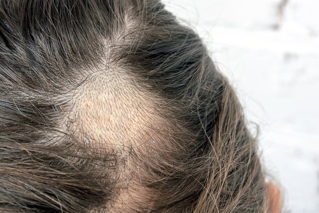
Tinea capitis is an infection caused by dermatophyte fungi, which are those that have a greater affinity for keratin, reaching the scalp and causing hair breakage and the appearance of one or more areas without hair on the head.
Tinea capitis is also known as tinea capitis or scalp and is mainly caused by fungi of the genus Trichophyton and Microsporum, which must be identified by the dermatologist to confirm the diagnosis of scalp ringworm and initiate the best treatment. See more about scalp ringworm.
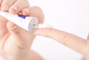
The treatment aims to combat the fungus and, thus, promote hair growth, and the doctor normally recommends the use of antimycotic remedies in the form of an ointment or tablet and the use of an appropriate shampoo. In cases where there are inflammatory signs, the use of corticosteroids may also be indicated.

Symptoms of tinea capitis
The main symptoms of tinea capitis are:
- Rounded plaque on the scalp without hair or with broken strands;
- The plaque may be single and large or several and small, depending on the infectious agent;
- Mild pain or redness in the area;
- Local peeling, in some cases;
- Presence of several crusts on the scalp, in some cases.
Furthermore, in some cases of Tinea capitis it is possible to notice the presence of inflammatory signs, such as swelling, blisters and secretion, this situation being known as kerion.
It is important that the dermatologist is consulted as soon as signs and symptoms possibly indicative of Tinea capitis are noticed, as this makes it possible to make the diagnosis, identify the infectious agent and initiate the most appropriate treatment.
Main causes
Tinea capitis is caused by dermatophyte fungi, that is, by fungi that have a greater affinity for keratin, in this case the keratin present in the hair. The main fungi associated with this disease are those of the genus Trichophyton, such as T. shearingo T. violaceum and the T. schoenleiniiand the genus Microsporum, mainly the M. canis.
How the diagnosis is made
The diagnosis of tinea capitis is made by the dermatologist initially by evaluating the characteristics of the hairless areas on the scalp.
Normally, the hairs are collected and the hairless plaques are scraped, which are sent to the laboratory for diagnosis to be made. Cultural and direct examinations can be carried out, in which the characteristics of the fungus are observed microscopically, and molecular biology techniques are used. to identify the fungus responsible for the symptoms.
In some cases, the doctor may also place UV light in the area where the collection will be carried out to identify the hairs that are emitting fluorescence, as this is a sign of the presence of the fungus, this examination being known as the Wood’s Lamp examination. Furthermore, according to the color emitted after exposure to UV light, the doctor can now identify the fungus and, thus, begin treatment immediately. See more details about Wood’s lamp.
What is the treatment like?
Treatment for Tinea capitis must be carried out in accordance with the dermatologist’s guidance and aims to eliminate the fungus responsible for the symptoms, and the use of antimycotic ointments or oral medications may be indicated.
In the case of children, the use of terbinafine or griseofulvin may be indicated, while for adults, the use of oral terbinafine or itraconazole is normally indicated. In addition, the doctor may also recommend the use of shampoo containing selenium sulfide, which can also help fight the fungus more quickly.
In cases where inflammatory signs are identified, in addition to antimycotic medication, the doctor may recommend the use of corticosteroid medications to alleviate signs of inflammation and promote healing.

Bibliography
- RODRIGUES, Gustavo S.; OLIVEIRA, Flávio M.; PEREIRA, Eduardo F.; CRUZ, Rosana CB Adult tinea capitis caused by Trichophyton violaceum in Brazil: case report and literature review. An Bras Dermatol. Vol 83. 5 ed; 544-548, 2008
- SILVA, CAROLINNE S.; NEUFELD, PAULO MURILLO; GOUVÊA, EMANUELA H.; ABREU, PAULA A. Etiology and epidemiology of tinea capitis: case series report and literature review. 2018. Available at: <http://www.rbac.org.br/artigos/etiologia-e-epidemiologia-da-tinea-capitis-relato-de-serie-de-casos-e-revisao-da-literatura/ >. Accessed on September 27, 2021
- BRAZILIAN SOCIETY OF DERMATOLOGY – RIO DE JANEIRO REGION. Do you know what tinea capitis is?. Available at: <https://sbdrj.org.br/34440/>. Accessed on September 27, 2021
- BARER, Michael R. et al. Medical Microbiology – A guide to Microbial Infections: Pathogenesis, immunity, laboratory investigation and control. 19 ed. Elsevier, 2018. 582-585.

Sign up for our newsletter and stay up to date with exclusive news
that can transform your routine!
Warning: Undefined array key "title" in /home/storelat/public_html/wp-content/plugins/link-whisper-premium/templates/frontend/related-posts.php on line 12
Warning: Undefined array key "title_tag" in /home/storelat/public_html/wp-content/plugins/link-whisper-premium/templates/frontend/related-posts.php on line 13