Retinal mapping, also known as fundus examination or fundoscopy, is an examination in which the ophthalmologist can observe the eye gel (vitreous humor), the optic nerve, blood vessels and the tissue of the eye responsible for capturing images. , being able to detect changes and allow the indication of treatment.
In addition, retinal mapping may also be indicated in premature babies, aged 32 weeks or less, or weighing 1,500 g or less, to assess the presence of retinopathy of prematurity, a common change in premature babies who It can lead to irreversible damage if not treated quickly. Understand better what retinopathy of prematurity is and how it should be treated.
Retinal mapping is carried out free of charge by the SUS, when indicated, however, it can also be carried out in private clinics, for a price that varies according to the location and clinic where the examination is carried out.

What is it for
Retinal mapping is indicated to identify injuries and changes caused by:
- Eye diseasessuch as glaucoma, retinal detachment, tumor, inflammation, lack of blood flow or medication poisoning, for example;
- Systemic diseases that cause eye damagesuch as diabetes, high blood pressure, rheumatic diseases, neurological diseases or blood diseases.
Furthermore, retinal mapping is an exam that can be done whenever there is a change in vision that cannot be corrected by wearing glasses.
The exam is part of the routine eye exam, but it also serves as a tool to diagnose problems that are leading to a decrease in vision, even with the best possible correction, whether with glasses or contact lenses, for example.
How is done
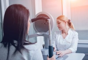
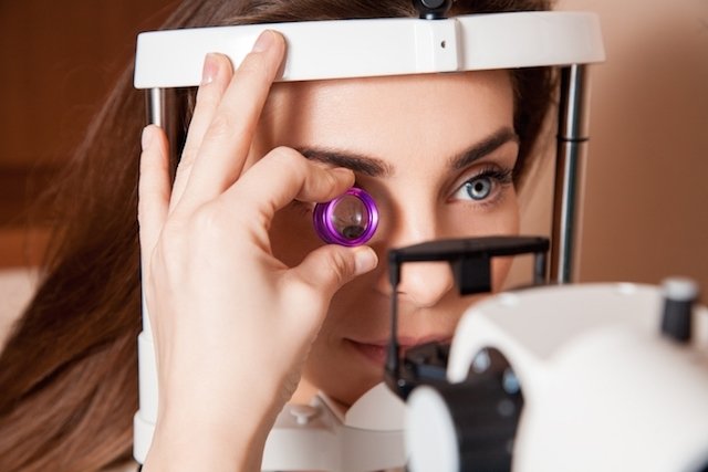
Retinal mapping is an exam carried out during a consultation with an ophthalmologist, which does not cause pain. To carry it out, a device called an indirect binocular ophthalmoscope and accessory lenses are used. Then, a beam of light is projected into the back of the eye, allowing the doctor to observe the region.
With this observation, the ophthalmologist will be able to identify possible changes and, if necessary, order other tests, such as tomography, or even recommend treatments, such as medicines to treat inflammation or surgery to reposition retinal detachment, for example.
To carry out the exam, the doctor may recommend dilation of the pupil, carried out with eye drops also applied during the consultation, shortly before the exam, which is why it is recommended to have a companion to help with the return home.
See also other eye exams that can be done to avoid vision complications.
Who should take the exam
Retinal mapping should be done more frequently in cases where there is a greater chance of having some changes in the retina, such as:
- People over 50 years old;
- Patients with chronic diseases such as high blood pressure, diabetes or rheumatological diseases;
- People with myopia;
- People who use medicines that can be toxic to the retina, such as chloroquine, chlorpromazine, tamoxifen or isotretinoin;
- Family or personal history of retinal detachment;
- After trauma or eye injuries.
With retinal mapping, it is possible to detect the main changes in the retina or eye diseases in general early, so that treatment can be carried out quickly, avoiding complications, such as loss of vision.

Sign up for our newsletter and stay up to date with exclusive news
that can transform your routine!
Warning: Undefined array key "title" in /home/storelat/public_html/wp-content/plugins/link-whisper-premium/templates/frontend/related-posts.php on line 12
Warning: Undefined array key "title_tag" in /home/storelat/public_html/wp-content/plugins/link-whisper-premium/templates/frontend/related-posts.php on line 13