Polyhydramnios corresponds to an increase in the volume of amniotic fluid, which generally happens due to increased production of this fluid, as a consequence of gestational diabetes, or due to the baby’s inability to absorb and swallow the liquid in normal quantities.
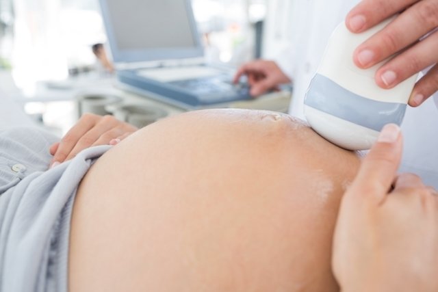
In most cases, polyhydramnios resolves on its own, however it is important that the obstetrician perform a morphological ultrasound, in order to observe whether the baby is developing normally, in addition to checking the total volume of amniotic fluid. If the amount is very excessive, the doctor may recommend draining the liquid to avoid possible risks to the mother or baby.
How to identify
Increased amniotic fluid can only be confirmed by the doctor through an ultrasound, however the woman may present some signs and symptoms that may be indicative of polyhydramnios, such as:
- Abdominal discomfort;
- Difficulty breathing;
- Smooth and shiny skin in the abdominal region;
- Swelling in the lower abdomen.
Furthermore, during a physical examination, the doctor may have difficulty palpating the fetus, and it may also be difficult to hear the fetal heartbeat.
From the moment it is verified that the amniotic fluid is increased, the doctor must assess the total volume of fluid and the severity of the symptoms, in addition to performing a morphological ultrasound to evaluate the baby’s characteristics, and request tests to investigate the presence of Gestational diabetes.
Main causes
Increased amniotic fluid can occur due to situations related to the woman or the baby, which cause greater fluid production or changes in the baby’s consumption. Thus, the main causes of polyhydramnios are:
- Gestational diabetes: the increase in the amount of sugar in the pregnant woman’s blood causes the baby to produce more urine, increasing the amount of amniotic fluid;
- Gastrointestinal problems in the baby: may decrease the baby’s ability to absorb amniotic fluid;
- Abnormal growth of blood vessels in the placenta: promotes an exaggerated production of amniotic fluid;
- Infections in pregnant women or babies such as rubella, cytomegalovirus, toxoplasmosis or syphilis;
- Chromosomal diseases such as Down Syndrome or Edwards Syndrome;
- Fetal malformationssuch as anencephaly, esophageal or duodenal atresia, diaphragmatic hernia;
- Anemia fetalin which there is a progressive decrease in the number of red blood cells and, consequently, hemoglobin in the developing baby’s blood.
The increase in the volume of amniotic fluid is normal between weeks 34 and 36 of pregnancy, with a progressive decrease as delivery approaches. However, if the amniotic fluid continues to increase, it is important to consult the obstetrician to assess the need to take any measures.
Possible complications of polyhydramnios
The increase in amniotic fluid can favor the appearance of some complications during pregnancy such as excessive fetal growth and development, placental abruption, premature birth, prolonged labor and difficulty breathing in women.
Generally, the earlier the increase in amniotic fluid appears during pregnancy and the more serious the problem, the greater the risk of developing consequences.
How the treatment is carried out
Treatment for increased amniotic fluid is usually not necessary, and it is only recommended to have regular appointments with the obstetrician to assess the amount of amniotic fluid over time. However, when the problem is caused by a disease, such as gestational diabetes, the doctor may recommend treatment for this problem in order to control the production of amniotic fluid.
In more serious cases in which the increase in amniotic fluid is causing difficulty breathing, severe abdominal pain or premature labor, drainage of the amniotic fluid may be recommended, this procedure being called amniodrainage. This procedure is very similar to amniocentesis, in which a thin needle is inserted into the abdomen to collect excess amniotic fluid. Understand better how amniocentesis is performed.

Bibliography
- GARRIDO, Adriana G.; FILHO, Evaldo TSS; NETTO, José Paulo S.; FERREIA, Adilson C. Ultrasound assessment of amniotic fluid: techniques and reference values. FEMALE. Vol 47. 1 ed; 46-51, 2019
- HEALTH DEPARTMENT OF THE STATE OF CEARÁ. Obstetrics Protocols. 2014. Available at: <https://www.saude.ce.gov.br/wp-content/uploads/sites/9/2018/06/protocolos_obstetricia_sesa_ce_2014_.pdf>. Accessed on March 4, 2021
- AQUINO, MARCELO A.; GRIIJÓ, MAURÍCIO. Amniotic Fluid Volume Changes. 2016. Available at: <http://www2.ebserh.gov.br/documents/215335/4407336/Protocolo+Alteracoes+Liquido+Amniotico.pdf/9fbd3b7a-8c9a-4708-b9c9-14ce9618b45a>. Accessed on March 4, 2021

Sign up for our newsletter and stay up to date with exclusive news
that can transform your routine!
Warning: Undefined array key "title" in /home/storelat/public_html/wp-content/plugins/link-whisper-premium/templates/frontend/related-posts.php on line 12
Warning: Undefined array key "title_tag" in /home/storelat/public_html/wp-content/plugins/link-whisper-premium/templates/frontend/related-posts.php on line 13