Obstetric ultrasound is an exam indicated to monitor pregnancy, assess the baby’s health, check the location of the placenta and calculate gestational age, and can also be used to assist in exams such as amniocentesis.
This exam is part of the prenatal routine, and is normally indicated between 11 and 14 weeks of pregnancy and repeated during pregnancy to monitor the baby’s development. Find out about other tests recommended during pregnancy.
Monitoring the pregnancy is important to identify and treat diseases that can affect the health of the baby and the mother, and it is recommended to regularly consult the obstetrician and carry out tests as recommended.

Obstetric ultrasound with color doppler
Obstetric ultrasound with color Doppler is indicated to evaluate blood flow through the baby’s umbilical artery and other vessels. This exam allows you to visualize blood circulation in color and identify possible changes. See how Doppler ultrasound is performed.
What is it for
Obstetric ultrasound is indicated for:
- Confirm suspected pregnancyespecially when the pregnancy test is positive and the symptoms are unclear;
- Check baby’s locationin case of suspected ectopic pregnancy;
- Estimate gestation timewhich is the gestational age;
- Track baby’s growth through specific measures;
- Identify changes in fetal development indicative of malformations and genetic syndromes;
- Assess the baby’s healthin case of accidents or vaginal bleeding, for example;
- Check the location and development of the placentawhich may indicate abnormalities such as placenta previa or accreta.
In addition, obstetric ultrasound is also normally used to guide other tests during pregnancy, such as amniocentesis, a test generally indicated to identify genetic changes in the baby. Check out what amniocentesis is and what it is for.
Does an obstetric ultrasound show the baby’s sex?
Obstetric ultrasound can show the baby’s sex, especially from the second trimester of pregnancy. Before then, it is more difficult to accurately identify gender through ultrasound examination. Find out when you can find out the baby’s sex.
Difference Between Obstetric and Morphological Ultrasound
Obstetric ultrasound is an exam used to monitor pregnancy, providing general information about the conditions of the pregnancy, the baby’s development and characteristics of the placenta, for example.
Morphological ultrasound is an exam mainly indicated to identify possible malformations in the baby and abnormalities in the development of the placenta, and is carried out in the 2nd trimester of pregnancy, between 18 and 22 weeks. Understand better what morphological ultrasound is and what it is for.
How is done
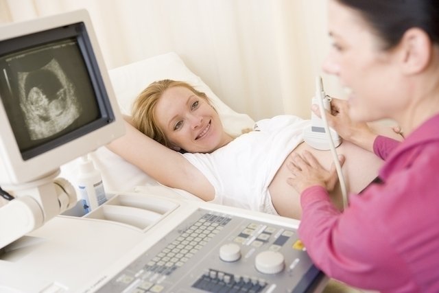
Obstetric ultrasound is performed on the belly or vagina, using a device called a transducer, which is connected to an ultrasound machine, and takes around 30 minutes.
When the examination is performed on the belly, the woman normally remains lying down with her abdomen exposed and the doctor or technician applies a gel to the belly and moves the transducer over the skin to obtain the images.
When it is done through the vagina, the woman usually puts on a type of hospital robe and needs to lie down with her legs supported, with the transducer placed in the vagina.
When to have the first obstetric ultrasound
The first obstetric ultrasound should be done between 11 and 14 weeks of pregnancy, being especially important to check the gestational age, and can be done through the belly or vagina, according to the obstetrician’s recommendations. Learn about the problems that the first ultrasound during pregnancy can detect.

Sign up for our newsletter and stay up to date with exclusive news
that can transform your routine!
Warning: Undefined array key "title" in /home/storelat/public_html/wp-content/plugins/link-whisper-premium/templates/frontend/related-posts.php on line 12
Warning: Undefined array key "title_tag" in /home/storelat/public_html/wp-content/plugins/link-whisper-premium/templates/frontend/related-posts.php on line 13