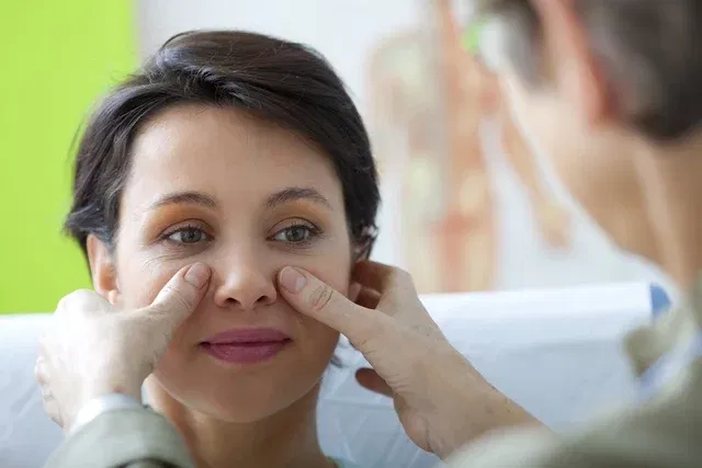
Nasofibroscopy is an exam that allows you to visualize the nasal cavity in more detail, which is done through the use of a device called a nasofibroscope, which has a camera that allows you to visualize the inside of the nose and the structures in this region, and record the images on a computer.
This exam is indicated to assist in the diagnosis of changes in the nasal cavity, such as deviations in the nasal septum, sinusitis, nasal tumors, among others, as it allows the identification of anatomical structures with precision and the visualization of the nasal cavity with a viewing angle and adequate lighting.
Therefore, it is important to consult an otorhinolaryngologist to assess the need for nasofibroscopy so that the diagnosis can be made and treatment indicated according to the condition.
What is it for
The main indications for nasofibroscopy are:
- Deviated nasal septum;
- Hypertrophy of the inferior turbinates or adenoids;
- Rhinitis and sinusitis;
- Lesions or tumors in the nose and/or throat;
- Snoring and sleep apnea;
- Changes in smell and/or taste;
- Nosebleeds;
- Frequent headache;
- Hoarseness;
- Frequent and constant coughing.
In addition, the otorhinolaryngologist may also recommend carrying out the examination with the aim of identifying the presence of foreign bodies in the upper airways.
Make an appointment with the nearest doctor for an assessment to be carried out and whether the exam is necessary:
Taking care of your health has never been easier!
Exam preparation
It is recommended that the person fast for 2 hours before undergoing the nasofibroscopy to avoid nausea and vomiting during the exam.
How the nasofibroscopy exam is performed
The nasofibroscopy exam is simple and quick, lasting around 15 minutes. To evaluate the nasal cavity, the otolaryngologist inserts a tube, known as a nasofibroscopic tube, which contains a small camera at its end. Thus, through this tube, the doctor can visualize the interior of the nose and nearby structures in more detail.
Anesthesia is not necessary to carry out the examination, however, as it may cause some discomfort, the doctor may recommend the application of local anesthesia to facilitate the examination.
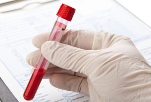
In some cases, when changes are seen in the nasal cavity, the doctor may take a small sample from the area so that a biopsy can be performed.
Bibliography
- DRHENRIQUEFURLAN. What is nasofibroscopy?. 2018. Available at: <http://www.drhenriquefurlan.com/v1/wp-content/uploads/2018/06/nasofibrescola.pdf>. Accessed on October 21, 2019
- FOIM; Angela BF et. at the.. Nasofibroscopy for the diagnosis of allergic rhinitis in children and adolescents. Brazilian Journal of Allergy and Immunopathology. 24. 6; 220-228, 2001

Sign up for our newsletter and stay up to date with exclusive news
that can transform your routine!
Warning: Undefined array key "title" in /home/storelat/public_html/wp-content/plugins/link-whisper-premium/templates/frontend/related-posts.php on line 12
Warning: Undefined array key "title_tag" in /home/storelat/public_html/wp-content/plugins/link-whisper-premium/templates/frontend/related-posts.php on line 13