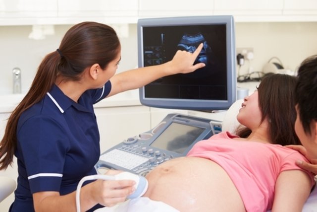
Morphological ultrasound is an imaging test that allows you to visualize the baby inside the uterus and all its organs, facilitating the identification of some diseases or malformations, such as Down syndrome or congenital heart defects, for example.
This exam, also known as morphological ultrasound or morphological USG, is normally done in the first trimester, between the 11th and 13th weeks of pregnancy, or in the second trimester, between the 20th and 24th weeks of pregnancy.
Morphological USG is part of prenatal exams, and marks the first moment in which parents are able to see the developing baby in detail. Know what other tests should be done during the second trimester of pregnancy.

What is it for
Morphological ultrasound is used to:
- Confirm the baby’s gestational age;
- Assess the baby’s size by measuring the head, chest, abdomen and femur;
- Assess the baby’s growth and development;
- Monitor the baby’s heartbeat;
- Check the position of the placenta, umbilical cord and amniotic fluid volume;
- Show abnormalities in the baby and possible illnesses or malformations.
Morphological ultrasound allows you to identify the baby’s developmental phase, as well as evaluate possible changes in the developmental phases.
In addition, this exam allows you to observe the development or changes in organs, such as the face, skull, brain, heart, lungs, stomach, abdominal wall, bladder, kidneys, bladder, arms, legs, hands, feet and spine.
When the baby has its legs apart, the doctor may also be able to observe the sex, which can then be confirmed with blood tests, for example. Check out a list of available techniques to try to identify the baby’s sex.
Schedule the morphological ultrasound in the closest region:
Taking care of your health has never been easier!
When to do morphological ultrasound
It is recommended to have a morphological ultrasound in the second trimester, between 20 and 24 weeks of gestation, as this is when the baby is already sufficiently developed.
However, this ultrasound can also be done in the first trimester, between the 11th and 13th week of pregnancy, but as the baby is not yet well developed, the results may not be as satisfactory.
Morphological ultrasound can also be done in the 3rd trimester, between 33 and 34 weeks of gestation, but this generally only happens when the pregnant woman did not undergo USG in the 1st or 2nd trimester, there is a suspicion of malformation in the baby or when the pregnant woman developed an infection that could harm the baby’s development.
In addition to morphological ultrasound, 3D and 4D ultrasounds show details of the baby’s face and also identify diseases. See when to do 3D and 4D ultrasound.
What diseases can be identified
Morphological ultrasound performed in the 2nd trimester can help identify several problems in the baby’s development, such as:
- Spina bifida;
- Anencephaly or hydrocephalus;
- Diaphragmatic hernia or gastroschisis;
- Cleft palate;
- Changes in the kidneys;
- Down, Edwards or Patau syndrome;
- Heart diseases.
In this way, morphological USG helps to evaluate the development of the fetus, and help identify the risk of malformations or diseases. See what a baby’s normal development should be like at 20 weeks.
How to prepare for an ultrasound
Normally, no special preparation is necessary to perform a morphological ultrasound.
However, as a full bladder can help improve images and also elevate the uterus, the obstetrician may advise drinking water before the exam, as well as avoiding completely emptying the bladder if you feel like going to the bathroom.
How is done
Morphological ultrasound is performed by the obstetrician, in specialized clinics, lasting around 20 to 60 minutes.
The ultrasound is performed with the woman lying on a stretcher and then a thin layer of gel is placed on the skin of the belly and then the transducer is positioned on top of this gel, sliding the device over the skin.
This device will generate images that can be viewed on a computer and must be analyzed by the doctor.
After finishing the exam, the doctor removes the gel with a paper towel and the woman can go home.
How to understand the result
The result of the morphological ultrasound examination must be evaluated by the obstetrician, according to the gestational age, through the evaluation of the organs, the main ones being:
1. Skull and central nervous system
Morphological USg allows evaluating the development of the skull and the central nervous system, through the measurement of several parameters, such as:
In addition, this exam evaluates the Lateral Ventricle (VL)/Cerebral Hemisphere (HC) relationship, which assesses the normal size of the ventricles and the presence of ventriculomegaly, which are ventricles with an increased size.
2. Face
Facial parameters are also evaluated in morphological US, the main ones being
In addition, this exam evaluates the DIO/DBP ratio (intraorbital distance/biparietal distance) and the nose/nasal bone (ON) ratio.
These parameters of the morphological examination of the face make it possible to evaluate changes such as anophthalmia, microphthalmia, hypo- and hypertelorism, which may be signs of syndromes.
3. Abdominal cavity
Morphological USG of the abdominal cavity generally evaluates the following parameters:
Furthermore, the morphological USG of the abdominal cavity allows the evaluation of organs such as the liver, spleen and kidneys, checking whether they are developing normally or whether they have changes.
4. Thoracic cavity
Morphological USG also evaluates parameters of the thoracic cavity, such as:
In addition, this exam evaluates the volume of the lungs and the size of the chest, in addition to the presence of lung or heart diseases.
5. Bones, spine and neck
USG allows you to evaluate the size and shape of the bones of the legs, arms, hands and feet, as well as the presence of bone fractures.
Furthermore, it also allows you to evaluate the spine and the presence of changes.
USG also evaluates the shape of the neck and the nuchal fold, which may be altered in some syndromes, such as Down syndrome.
6. Fetal attachments
Fetal appendages are also evaluated on morphological USG, such as:
In addition to these parameters, morphological USG also allows estimating the expected date of delivery (DPP).

Sign up for our newsletter and stay up to date with exclusive news
that can transform your routine!
Warning: Undefined array key "title" in /home/storelat/public_html/wp-content/plugins/link-whisper-premium/templates/frontend/related-posts.php on line 12
Warning: Undefined array key "title_tag" in /home/storelat/public_html/wp-content/plugins/link-whisper-premium/templates/frontend/related-posts.php on line 13