The echocardiogram is a diagnostic test that allows the assessment, in real time, of the heart’s movements, as well as some characteristics, such as heart size, shape of the valves, muscle thickness and the heart’s ability to function, in addition to blood flow.
This exam is carried out using an ultrasound with high frequency sound waves, also allowing us to see the condition of the large vessels of the heart, pulmonary artery and aorta, at the time the exam is being carried out.
The echocardiogram, also called echocardiography or ultrasound of the heart, has different types, such as one-dimensional, two-dimensional and Doppler, which are requested by the doctor according to what they want to evaluate, and can be performed free of charge by the SUS or carried out in clinics or private hospitals.
What is it for
Echocardiography is indicated for:
- Analyze cardiac function;
- Assess the size and thickness of the heart walls;
- Evaluate the structure of the valves, valve malformations and visualization of blood flow;
- Calculate cardiac output, which is the amount of blood pumped per minute;
- Check changes and diagnose diseases in the membrane that covers the heart, called the pericardium;
- Investigate the cause of symptoms, such as heart palpitations, shortness of breath, excessive tiredness or fainting;
- Diagnose heart diseases, such as heart murmur, hypertrophic cardiomyopathy, thrombi in the heart, aneurysm or aortic dissection;
- Identify congenital heart malformations;
- Investigate masses and tumors in the heart;
- Monitor the evolution of heart diseases, such as heart failure or heart valve diseases;
- Assess the heart in cases of suspected coronary artery disease, pulmonary embolism, hypotension, respiratory failure of unknown cause or pericardial effusion or complications from cardiothoracic surgery.
The echocardiogram is a simple test used to evaluate the functioning of the heart in people with or without cardiac symptoms, or who have chronic cardiovascular diseases, such as hypertension or diabetes.
Types of echocardiogram
Depending on the objective of carrying out the exam, different echocardiograms can be performed, the main ones being:
- Transthoracic echocardiogram: it is the most commonly performed exam, in which the equipment is placed in the thoracic region;
- Stress echocardiogram: it is performed in the same way as the transthoracic echocardiogram, however, the images are obtained before and after performing exercises, or using medication, in a hospital environment;
- Fetal echocardiogram: carried out during pregnancy to evaluate the baby’s heart and identify diseases;
- Doppler echocardiogram: especially indicated for evaluating blood flow through the heart, especially useful in valvular heart disease;
- Transesophageal echocardiogram: It is also indicated to evaluate the esophagus region in search of diseases.
This exam can also be carried out in a one-dimensional or two-dimensional way, which means that the images generated evaluate 2 different angles at the same time, and in a three-dimensional way, which evaluates 3 dimensions at the same time, being more modern and credible.
How to prepare
The echocardiogram is a simple procedure that does not require preparation, with the exception of the transesophageal echocardiogram, in which it is recommended that the person not eat for approximately 3 hours before the exam.
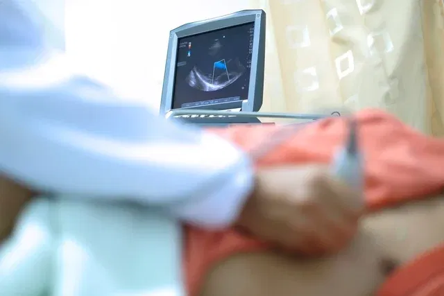
How is an echocardiogram performed?
The echocardiogram is typically done in the cardiologist’s office or at an imaging clinic, and lasts 15 to 20 minutes. The person simply needs to lie on the stretcher on their back or on their left side, and remove or pull up their shirt.
Then, the doctor applies some gel to the area of the chest where the heart is located and slides the ultrasound equipment that generates images onto a computer, from several different angles.
During the exam, the doctor may ask the person to change position or perform specific breathing movements so that a more accurate result can be obtained. After the echocardiogram, the person can return to their normal routine.
Who shouldn’t do
There is no contraindication for carrying out this test, which can be carried out even on babies and children, and is also recommended for amateur and professional athletes and sedentary people who are about to start practicing physical activity.
However, transesophageal echocardiography is not indicated in cases of narrowing of the esophagus, tumors, acute gastrointestinal bleeding or diverticula.
Stress echocardiography should not be performed in cases of recent heart attack, narrowing of the aorta with severe symptoms, exacerbation of heart failure, uncontrolled arrhythmias, unstabilized unstable angina or acute pericarditis, for example.
Bibliography
- LEE, J.; et al. Interventional echocardiography: Opportunities and challenges in an emerging field. Echocardiography. 39. 7; 975-984, 2022
- VIEILLARD-BARON, A.; et al. A decade of progress in critical care echocardiography: a narrative review. Intensive Care Med. 45. 6; 770-788, 2019
- DEPLA, AL; et al. Effect of maternal diabetes on fetal heart function on echocardiography: systematic review and meta-analysis. Ultrasound Obstet Gynecol. 57. 4; 539-550, 2021
- BELFRAGE, K.; et al. Initial fetal to initial postnatal echocardiogram in uncomplicated atrioventricular septal defects: Do significant changes occur?. Echocardiography. 37. 12; 2102-2106, 2020
- ZHANG, J.; et al. Fully Automated Echocardiogram Interpretation in Clinical Practice. Circulation. 138. 16; 1623-1635, 2018
- FITZSIMONS, S.; DOUGHTY, R. N. Role of transthoracic echocardiogram in acute heart failure. Rev Cardiovasc Med. 22. 3; 741-754, 2021
- Yu, S.; et al. Transesophageal Echocardiogram to the Rescue in Diagnosing Ascending Aortic Pseudoaneurysm. Anesthesiology. 132. 1; 158, 2020
- OMEROVIC, S.; JAIN, A. IN: STATPEARLS (INTERNET). TREASURE ISLAND (FL): STATPEARLS PUBLISHING. Echocardiogram. 2022. Available at: <https://www.ncbi.nlm.nih.gov/books/NBK558940/>. Accessed on March 20, 2023
- AMERICAN HEART ASSOCIATION. Echocardiogram (Echo). Disponível em: <https://www.heart.org/en/health-topics/heart-attack/diagnosing-a-heart-attack/echocardiogram-echo>. Acesso em 29 jun 2021
- NHS. Echocardiogram. Available at: <https://www.nhs.uk/conditions/echocardiogram/>. Accessed on June 29, 2021

Sign up for our newsletter and stay up to date with exclusive news
that can transform your routine!
Warning: Undefined array key "title" in /home/storelat/public_html/wp-content/plugins/link-whisper-premium/templates/frontend/related-posts.php on line 12
Warning: Undefined array key "title_tag" in /home/storelat/public_html/wp-content/plugins/link-whisper-premium/templates/frontend/related-posts.php on line 13