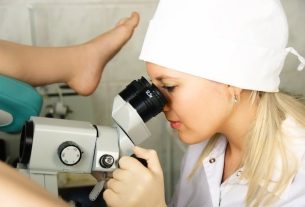
Dermoscopy is a type of non-invasive dermatological examination that aims to analyze the skin in more detail, being useful in the investigation and diagnosis of changes, such as skin cancer, keratosis, hemangioma and dermatofibroma, for example.
This detailed analysis is possible through the use of a device, a dermatoscope, which shines a light on the skin and has a lens that allows the skin to be observed in more detail, as it has a magnification power of around 6 to 400 times the actual size.
After performing a dermoscopy, the dermatologist is able to evaluate skin changes in more detail, making it possible to recommend the most appropriate treatment for the situation, if necessary.
What is it for
Dermoscopy is used to evaluate in more detail changes in the skin that may be suggestive of malignancy, such as:
- Spots on the skin that may be suggestive of melanoma;
- Seborrheic keratosis;
- Hemangioma;
- Dermatofibroma;
- Signals;
- Lesions possibly caused by infections, as in the case of leishmaniasis and HPV
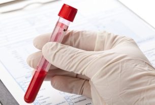
As dermoscopy promotes skin enlargement, in some cases, especially in cases where the presence of pigmented lesions is verified, the severity of the change and the presence of infiltrations can be observed. Thus, the doctor can recommend early treatment for the situation while waiting for the results of other tests that may have been requested, such as a skin biopsy, for example. See what it is for and how a skin biopsy is performed.
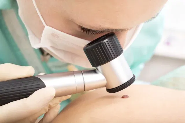
How it is made
Dermoscopy is a non-invasive examination carried out by a dermatologist, in which a device is used that allows the skin to be magnified up to 400x, making it possible to observe the innermost structure of the skin and make a more detailed assessment of the possible change. The device used is called a dermatoscope, it is placed directly on the lesion and emits a beam of light so that the lesions can be observed.
Dermatoscopia digital
Digital dermoscopy is similar to conventional dermoscopy, however the dermatoscope is connected to digital cameras or computers so that images can be collected during the examination, which will be stored and can be consulted by the dermatologist, helping to evaluate the evolution of the change.
Bibliography
- REGIONAL MEDICINE COUNCIL OF THE STATE OF PARANÁ. 2018. Available at: <https://sistemas.cfm.org.br/normas/arquivos/pareres/PR/2018/2700_2018.pdf>. Accessed on October 10, 2022
- REZZE, Gisele G.; SÁ, Bianca CS; NEVES, Rogério Izar. Dermoscopy: the pattern analysis method. An Bras Dermatol. 3 ed; 261-268, 2006
- JUNIOR, Walter Belda et. to the.. Dermatology treatise. 2. São Paulo: Atheneu, 2014. 99-106.

Sign up for our newsletter and stay up to date with exclusive news
that can transform your routine!
Warning: Undefined array key "title" in /home/storelat/public_html/wp-content/plugins/link-whisper-premium/templates/frontend/related-posts.php on line 12
Warning: Undefined array key "title_tag" in /home/storelat/public_html/wp-content/plugins/link-whisper-premium/templates/frontend/related-posts.php on line 13