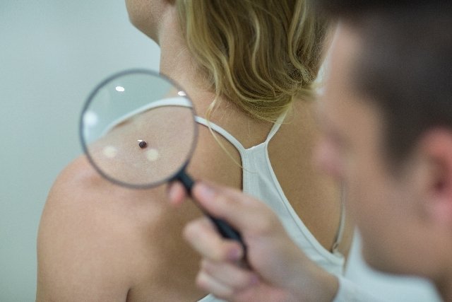
The dermatological examination is a simple and quick examination that aims to identify changes that may be present in the skin, and the examination must be carried out by the dermatologist in his office.
However, the dermatological examination can also be done at home and to do this, the person can stand in front of the mirror and carefully observe their body, looking for new signs, spots, scars, peeling or itching, including the back of the neck. of the ears and between the toes.
If new signs are observed, it is important to go to the dermatologist so that the examination can be carried out in more detail and the diagnosis can be made.

How is the dermatological examination performed?
The dermatological examination is simple, quick and does not require any type of preparation, because it consists of observing lesions, spots or signs present on the skin. This exam is normally requested for users of public swimming pools, private clubs and some gyms.
The exam is carried out in the dermatologist’s office and takes place in two stages:
- anamnesein which the doctor will ask questions about the injury, such as when it started, when the first symptom appeared, what the symptom is like (whether it itches, hurts or burns), whether the injury has spread to another part of the body and whether the injury has progressed.
- Physical examin which the doctor will observe the person and the lesion, paying attention to the characteristics of the lesion, such as color, consistency, type of lesion (plaque, nodule, spots, scar), shape (target, linear, rounded), arrangement ( grouped, scattered, isolated) and distribution of the lesion (localized or disseminated).
Through a simple dermatological examination, you can discover various diseases such as chilblains, chilblains, ringworm, herpes, psoriasis and other more serious diseases such as melanoma, which is a type of skin cancer that can easily spread to other organs. Learn how to identify melanoma.
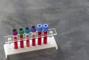
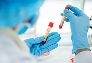
Auxiliary diagnostic tests
Some diagnostic tests can be used to complement the dermatological examination, when the physical examination is not sufficient to determine the cause of the lesion, they are:
- Biopsy, in which part of the injured region or sign is removed so that the characteristics can be evaluated and the diagnosis can be finalized. Biopsy is widely used to diagnose skin cancer, for example. See what the first signs of skin cancer are;
- Shaved, in which the doctor shaves the lesion so that it can be taken to the laboratory for analysis. This test is normally done to diagnose fungal infections;
- Wood’s Lightwhich is widely used to evaluate the spots present on the skin and make the differential diagnosis with other diseases through the fluorescence pattern, such as erythrasma, in which the lesion fluoresces in a bright red-orange tone, and vitiligo, which turns blue- bright;
- Tzanck cytodiagnosis, which is done to diagnose lesions caused by viruses, such as herpes, which usually manifests itself through blisters. Therefore, the material used to carry out this diagnostic examination are bubbles.
These exams help the dermatologist to define the cause of the lesion and establish the appropriate treatment for the patient.

Sign up for our newsletter and stay up to date with exclusive news
that can transform your routine!
Warning: Undefined array key "title" in /home/storelat/public_html/wp-content/plugins/link-whisper-premium/templates/frontend/related-posts.php on line 12
Warning: Undefined array key "title_tag" in /home/storelat/public_html/wp-content/plugins/link-whisper-premium/templates/frontend/related-posts.php on line 13