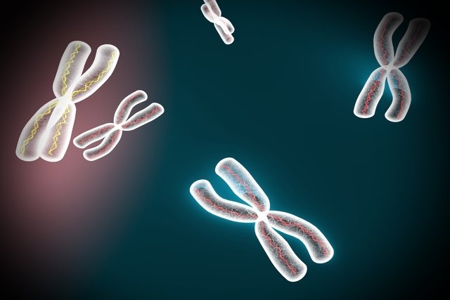
Cytogenetics is an exam that aims to analyze chromosomes and, thus, identify changes in the structure and number of chromosomes, and is generally indicated to diagnose genetic diseases or some types of cancer.
This test is done by collecting a sample of blood, tissue or bone marrow and can be done at any age, even during pregnancy to check for possible genetic changes in the baby.
Cytogenetics allows the doctor and patient to have an overview of the genome, helping the doctor to make the diagnosis and direct treatment, if necessary. This test does not require any preparation and the collection does not take long to be carried out, however the result may take between 3 and 10 days to be released according to the laboratory.

What is it for
The human cytogenetic examination is indicated to investigate possible chromosomal changes, both in children and adults, such as:
- Down syndrome;
- Edwards syndrome;
- Patau syndrome;
- DiGeorge Syndrome;
- Cri-Du-Chat Syndrome;
- Klinefelter syndrome;
- Hermaphroditism or pseudohermaphroditism;
- Leukemias;
- Lymphomas.
Generally, this test is indicated in cases of family history of some genetic alteration, changes in obstetric ultrasound, advanced maternal age, recurrent miscarriages or changes in biochemical tests.
Furthermore, the cytogenetic test in the case of cancer can help classify the type of leukemia, in addition to evaluating the best treatment, as well as the response to treatment, for example.
What the exam identifies
The cytogenetic exam allows you to identify changes in chromosomes, such as:
- Numerical changeswhich are characterized by an increase or decrease in the number of chromosomes, such as what happens in Down syndrome, in which the presence of three chromosomes 21 is verified, with the person having 47 chromosomes in total;
- Structural changeswhich are characterized by the substitution, exchange or elimination of a certain region of a chromosome, such as Cri-du-Chat syndrome, in which a piece of chromosome 5 is lost.
Thus, this test evaluates the chromosome, which is a structure made up of DNA and proteins that is distributed in cells in pairs, 23 pairs.
In this way, from the karyogram, which corresponds to the chromosome organization scheme, it is possible to identify changes in the number and structure of chromosomes.
How is done
The cytogenetic test is normally carried out using a blood sample, but it can also be carried out using tissue or bone marrow samples.
In the case of the examination in pregnant women, whose objective is to evaluate the fetus’ chromosomes, amniotic fluid, chorionic villi, or even small amounts of blood are collected.
After collecting the samples and sending them to the laboratory, the cells are cultivated so that they multiply and then a substance is added that inhibits cell division, causing the chromosome to be in its most condensed form and better visualization.
Types of cytogenetic exam
Depending on the objective of the examination, different molecular techniques can be applied to obtain information on the person’s karyotype, the most commonly used being:
1. G Banding
The G-banding technique, or karyotyping, consists of applying Giemsa dye, which allows visualization of the chromosomes.
This technique is very effective for detecting numerical, mainly structural, changes in the chromosome, being the main molecular technique applied in cytogenetics for diagnosing and confirming Down syndrome, for example, which is characterized by the presence of an extra chromosome.
2. FISH technique
The FISH technique is more specific and sensitive, being most used to assist in the diagnosis of cancer, as it allows the identification of small changes in chromosomes and rearrangements, in addition to also identifying numerical changes in chromosomes.
Despite being quite effective, the FISH technique is more expensive, as it uses fluorescently labeled DNA probes, requiring a device to capture the fluorescence and allow visualization of the chromosomes. Furthermore, there are more accessible techniques in molecular biology that allow the diagnosis of cancer.
Bibliography
- WAN, T. S. Cancer Cytogenetics: An Introduction. Methods Mol Biol. 1541. 1-10, 2017
- MONTAZERINEZHAD, S.; et al. Chromosomal abnormality, laboratory techniques, tools and databases in molecular Cytogenetics. Mol Biol Rep. 47. 11; 9055-9073, 2020
- NIH – NATIONAL CANCER INSTITUTE. Cytogenetics. Disponível em: <https://www.cancer.gov/publications/dictionaries/cancer-terms/def/cytogenetics>. Acesso em 24 abr 2023
- OZKAN, E.; LACERDA, M. P. IN: STATPEARLS (INTERNET). TREASURE ISLAND (FL): STATPEARLS PUBLISHING. Genetics, Cytogenetic Testing And Conventional Karyotype. 2022. Available at: <https://www.ncbi.nlm.nih.gov/books/NBK563293/>. Accessed on April 24, 2023
- KANNAN, T. P.; ZILFALIL, B. A. Cytogenetics: Past, Present And Future. Malays J Med Sci. 16. 2; 4–9, 2009

Sign up for our newsletter and stay up to date with exclusive news
that can transform your routine!
Warning: Undefined array key "title" in /home/storelat/public_html/wp-content/plugins/link-whisper-premium/templates/frontend/related-posts.php on line 12
Warning: Undefined array key "title_tag" in /home/storelat/public_html/wp-content/plugins/link-whisper-premium/templates/frontend/related-posts.php on line 13


