Cordocentesis, or fetal blood sample, is a prenatal diagnostic test, carried out from 18 or 20 weeks of gestation, which consists of taking a sample of the baby’s blood from the umbilical cord, to detect any chromosomal deficiency. in the baby, such as Down Syndrome, or diseases such as toxoplasmosis, rubella, fetal anemia or cytomegalovirus, for example.
The main difference between cordocentesis and amniocentesis, which are 2 prenatal diagnostic tests, is that Cordocentesis analyzes the baby’s umbilical cord blood, while Amniocentesis analyzes only the amniotic fluid. The karyotype result comes out in 2 or 3 days, which is one of the advantages over amniocentesis, which takes around 15 days.

When to do cordocentesis
Indications for cordocentesis include the diagnosis of Down Syndrome, when it cannot be obtained through amniocentesis, when ultrasound results are inconclusive.
Cordocentesis allows the study of DNA, karyotype and diseases such as:
- Blood diseases: Thalassemia and sickle cell anemia;
- Blood clotting diseases: Hemophilia, Von Willebrand Disease, Autoimmune Thrombocytopenia, Thrombocytopenic Purpura;
- Metabolic diseases such as Duchenne Muscular Dystrophy or Tay-Sachs Disease;
- To identify why the baby is growth restricted, and
- To identify hydrops fetalis, for example.
Furthermore, it is also very useful for diagnosing whether the baby has a congenital infection and can also be indicated as a form of treatment for intrauterine blood transfusion or when it is necessary to administer medications to treat fetal diseases, for example.
Find out other tests for diagnosing Down Syndrome.
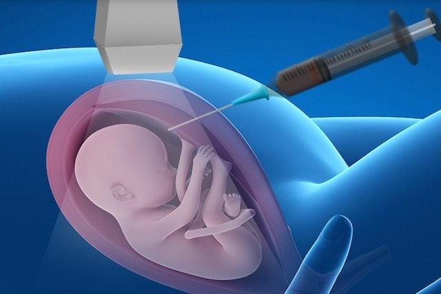
How cordocentesis is performed
No preparation is necessary before the exam, however, the woman must have undergone an ultrasound exam and a blood test before cordocentesis to indicate her blood type and RH factor. This exam can be carried out in the clinic or hospital, as follows:
- The pregnant woman lies face up;
- The doctor applies local anesthesia;
- With the help of ultrasound, the doctor inserts a needle more specifically into the place where the umbilical cord and placenta join;
- The doctor takes a small sample of the baby’s blood measuring around 2 to 5 ml;
- The sample is taken to the laboratory for analysis.
During the exam, the pregnant woman may feel abdominal cramps and should therefore rest for 24 to 48 hours after the exam and not have intimate contact for 7 days after cordocentesis.
After the exam, symptoms such as fluid loss, vaginal bleeding, contractions, fever and pain in the lower abdomen may appear. To relieve pain and discomfort, it may be useful to take a Buscopan tablet, under medical advice.
What are the risks of cordocentesis
Cordocentesis is a safe procedure but has risks, like any other invasive exam, and that is why the doctor only requests it when there are more advantages than risks for the mother or baby. The risks of cordocentesis are low and manageable, but include:
- About 1 risk of miscarriage;
- Blood loss at the site where the needle is inserted;
- Decrease in the baby’s heartbeat;
- Premature rupture of the membranes, which can favor premature birth.
Generally, the doctor orders a cordocentesis when there is a suspicion of a genetic syndrome or disease that has not been identified through amniocentesis or ultrasound.

Sign up for our newsletter and stay up to date with exclusive news
that can transform your routine!
Warning: Undefined array key "title" in /home/storelat/public_html/wp-content/plugins/link-whisper-premium/templates/frontend/related-posts.php on line 12
Warning: Undefined array key "title_tag" in /home/storelat/public_html/wp-content/plugins/link-whisper-premium/templates/frontend/related-posts.php on line 13