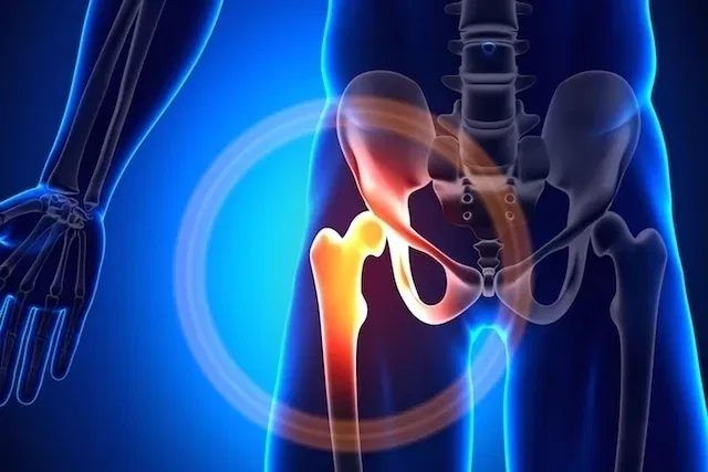
Congenital short femur is a bone malformation characterized by the reduction in size or absence of the femur, which is the thigh bone and the largest bone in the body. This change may occur as a result of the use of medication during pregnancy or a viral infection, however the causes of this malformation are not yet fully understood.
A congenital short femur can be identified during pregnancy, from the second trimester onwards, through an ultrasound examination, and can be indicative of diseases such as Down syndrome, dwarfism or achondroplasia, or simply be shortening of this bone. From the moment the diagnosis of short femur is made, the doctor can establish the treatment to be followed after the baby is born.
How to identify
The congenital short femur can be identified during pregnancy through an ultrasound performed during prenatal care, which measures the size of the femur, which varies according to gestational age.
The baby at 24 weeks is, on average, 42 mm, while at week 36 it is 69 mm and at week 40 of gestation, 74 mm. These measurements are approximate and therefore, in some cases, the baby He may be growing as expected even if his femur is smaller for his age, and it is important for the doctor to regularly monitor the baby’s development.
After identifying that the femur is smaller than it should be, the doctor must also observe what type of change the baby has, which could be:
- Type A: A small part of the femur below the head of the femur is deficient or absent;
- Type B: The head of the femur is attached to the lowest part of the bone;
- Type C: The head of the femur and the acetabulum, which is where the hip fits, are also affected;
- Type D: Most of the femur, acetabulum, and part of the hip are missing.
Often a small change is found at the end of pregnancy, but the height of the parents and the family must also be taken into account because if the parents are not very tall, their baby should not be either and this does not indicate any health problem. .
Furthermore, in some cases no changes are identified during pregnancy, only after birth through examinations carried out by the pediatrician, and changes in the length of the femur may be identified by the doctor due to the incorrect fit of this bone to the hip bone, characterizing congenital dysplasia. of the hip. Understand what congenital hip dysplasia is.
Possible causes
The causes of congenital short femur are not yet well understood, however it is believed that it may be related to infections during pregnancy, drug use and/or exposure to radiation during pregnancy.
Furthermore, the use of thalidomide, for example, could also favor the development of this change, because this medication is related to fetal malformations.
How the treatment is carried out
The treatment of congenital short femur is quite time-consuming, aims to improve the baby’s quality of life and must be guided by the pediatrician according to the type of shortening.
Furthermore, treatment is indicated according to the estimated size of the femur in adult life, and may be indicated in milder cases, where the shortening is up to 2 cm, the use of shoes with elevated soles or special insoles to compensate for the difference and prevent complications such as scoliosis, back pain and joint compensation, for example.
Other possible treatment indications for short femur are:
- For shortening between 2 and 5 cm in adults: surgery can be performed to cut the healthy leg bone so that they are the same size, surgery can be performed to lengthen the femoral or tibial bone, and while waiting for the ideal time for surgery, compensation can only be used with suitable footwear or a leg prosthesis;
- For shortening of more than 20 cm in adults: It may be necessary to amputate the leg and use a prosthesis or crutches for life. In this case, surgery is the most effective treatment and aims to add prostheses to the bone so that the person continues to walk normally. Surgery should preferably be performed before 3 years of age.
In any case, physiotherapy is always indicated to reduce pain, facilitate development and avoid muscle compensations or prepare for surgery, for example, but each case must be analyzed personally because physiotherapy treatment will be different for each person because each person’s needs may vary. not be those of the other.

Bibliography
- BORGES, Igor; DUARTE, Hannah Claudia S.; ÁVILA, Elizabeth M. et al. Congenital Short Femur: Case Report. RESU – HEALTH EDUCATION MAGAZINE. Vol 6. 1 ed; 2018
- FERNANDES, Henrique PA; BARRONOVO, Daniel Gabriel N.; RODRIGUES, Fábio Lucas; HONO, Marcos. Femoral bone lengthening with monoplanar external fixator associated with a locked intramedullary nail. Brazilian orthopedics journal. Vol 52. 1 ed; 82-86, 2017

Sign up for our newsletter and stay up to date with exclusive news
that can transform your routine!
Warning: Undefined array key "title" in /home/storelat/public_html/wp-content/plugins/link-whisper-premium/templates/frontend/related-posts.php on line 12
Warning: Undefined array key "title_tag" in /home/storelat/public_html/wp-content/plugins/link-whisper-premium/templates/frontend/related-posts.php on line 13