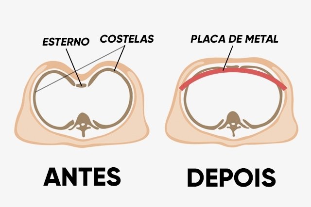
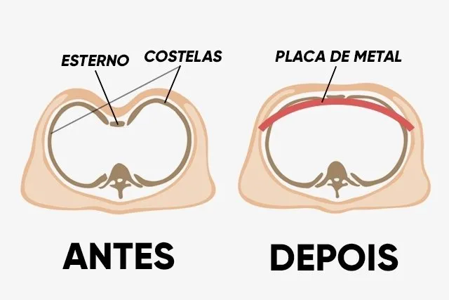
The excavated breast is a congenital malformation in which the bone of the sternum presents a depression in the center of the chest, in the region between the ribs, and can be identified by the neonatologist soon after birth.
This condition, also known as pectus excavatum, can cause an alteration of body image that, although it does not bring risk of life, can hinder the development of self-esteem or cause psychological changes, especially in the child.
The excavated breast can lead, in some cases, to compression of the organs of the region, which increases the risk of respiratory tract infections and difficulty breathing. In these situations, the orthopedist should be consulted who may indicate a surgery to reposition the bones in the correct place.
Symptoms of excavated breast
The main symptoms of excavated breast are:
- Depression in the middle of the chest, which can vary according to severity;
- Intolerance to exercise;
- pain in the chest;
- Fatigue;
- tonight;
- cardiac papitations;
- Wheezing when breathing and/or coughing;
- Recurrent respiratory infections.
These symptoms may appear in more severe situations of the excavated chest, as most people have only signs of depression in the chest between the ribs.
Although the excavated chest can be identified soon after birth, in many cases there is worsening as the child develops, so treatment is usually only indicated after adolescence, to reduce the risk of the problem reappearing.
How to confirm the diagnosis
The diagnosis of excavated breast can be made by the neonatologist soon after birth or by the pediatrician or orthopedist throughout the life of the child and until adolescence, being made through the physical evaluation of the chest.
Book an appointment with a pediatrician in the nearest area:
Taking care of your health has never been easier!
Make an appointment with our Pediatricians and receive the personalized care you deserve.
In addition, the doctor may indicate imaging tests, such as chest X-rays, CT scans or MRI, to assess whether there is any compression of the lungs or heart.
Other tests that may be ordered by the doctor are electrocardiogram, echocardiogram or pulmonary function tests, for example.
Possible causes
The cause of the appearance of the excavated breast is not known, however, it is more common in boys and people who have a family history of this malformation, and is usually related to the excessive growth of the cartilage that connects the ribs to the sternum bone.
This malformation can be observed in conditions such as Marfan syndrome, Noonan syndrome, Poland syndrome and osteogenesis imperfecta, for example.
How is the surgery done
The treatment of the excavated chest should be done by the pediatrician or orthopedist and is usually done with surgery to reposition the bones in the correct place in the most severe cases.
The excavated chest surgery can be done in two different ways, depending on the severity and age of the person. However, in both cases it is done with general anesthesia and it is necessary to stay hospitalized about 1 week.
The main types of surgery for the dugled chest are:
- Open or Ravitch surgery: it is used in adults, in moderate to severe cases, whose chest is rigid and very asymmetrical and lasts between 4 and 6 hours. In this technique a horizontal cut is made in the chest to remove the abnormal cartilage that connects the ribs to the bone of the sternum, allowing the bone to return to its correct position. Then surgical materials are placed to keep the chest in the correct position;
- Minimally invasive or Nuscive surgery: it is usually done in children and in mild to moderate cases and lasts between 1 and 2 hours. In this technique two small cuts are made under the armpit and then a metal bar is inserted between one cut and another, in order to push the sternum out to the correct position.
This is a very painful surgery and, therefore, after surgery, it is necessary to stay hospitalized especially to do analgesics directly in the vein and improve comfort, and the discharge is given as soon as the pain decreases and there are no complications.
How is the recovery
In the period after discharge, it is necessary to go to frequent consultations with the doctor to do X-ray tests or computed tomography in order to evaluate whether the sternum remains in the correct position. With these evaluations it is also possible to determine the best time to remove the surgical material or metal bar left during surgery.
In the case of open surgery, the material is usually removed after 6 to 12 months, while the bar of minimally invasive surgery is removed only after 2 or 3 years.
During this period it is also important to be aware of signs of infection or rejection of surgical material left in the body, such as swelling or redness at the site of the courts, fever above 38oC or excessive fatigue, for example.
Sports activities should only be started with approval of the doctor, avoiding those of greater impact and risk of injuries, such as football, basketball or martial arts.

Sign up for our newsletter and stay up to date with exclusive news
that can transform your routine!
Warning: Undefined array key "title" in /home/storelat/public_html/wp-content/plugins/link-whisper-premium/templates/frontend/related-posts.php on line 12
Warning: Undefined array key "title_tag" in /home/storelat/public_html/wp-content/plugins/link-whisper-premium/templates/frontend/related-posts.php on line 13