Cardiotocography is an exam carried out during pregnancy to check the baby’s heartbeat and well-being, and is carried out with sensors attached to the pregnant woman’s belly that collect this information, being especially recommended for pregnant women after 37 weeks or in periods close to the childbirth.
This exam can also be carried out during labor to monitor the health of the baby at this time, in addition to evaluating the woman’s uterine contractions.
The fetal cardiotocography exam must be carried out in clinics or obstetrics units, which have equipment and doctors prepared for the exam.

What is it for
Cardiotocography is an exam that serves to monitor the fetal heartbeat and to assess the well-being of the fetus, because it also monitors fetal movements and the baby’s oxygenation, being useful for preventing and diagnosing fetal distress, which is a situation in which the baby does not receive sufficient amounts of oxygen during pregnancy or birth. Learn more about fetal distress.
Furthermore, cardiotocography serves to monitor uterine contractions, but is not capable of measuring their intensity.
When is it indicated
Fetal cardiotocography may be indicated after 37 weeks only for a preventive assessment of the baby’s heartbeat. However, it may be indicated before week 37 of pregnancy, especially when the woman presents a risk situation, such as:
- Gestational diabetes;
- Uncontrolled high blood pressure;
- Pre eclampsia;
- Anemia grave;
- Heart, kidney or lung changes;
- Changes in the blood clotting process;
- Infections;
- The woman’s advanced age or young age.
Furthermore, cardiotocography may be indicated when there are conditions that may increase the risk for the woman and/or the baby at the time of birth, such as low amniotic fluid, changes in uterine contraction during childbirth, uterine bleeding, multiple pregnancy, abruption. of the placenta and long labor, for example.
In this way, by carrying out this exam, it is possible to intervene as quickly as possible, in case changes are noticed in the baby’s well-being, caused by suffocation, lack of oxygen, fatigue or arrhythmias, for example. EThis assessment can be carried out at different periods of pregnancy, such as:
- Non-antepartum: It is done at any time after 28 weeks of pregnancy, preferably after 37 weeks, to assess the baby’s heartbeat.
- No intraparto: in addition to the heartbeat, it evaluates the baby’s movements and the contractions of the mother’s uterus during childbirth.
The checks carried out during this exam are part of the set of fetal vitality assessments, as well as others such as Doppler ultrasound, which measures blood circulation in the placenta, and the fetal biophysical profile, which takes several measurements to observe the correct development of the baby. Find out more about the tests recommended for use in the third trimester of pregnancy.
How it is made
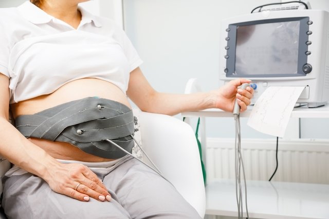
Cardiotocography is a simple and non-invasive exam and consists of placing electrodes with sensors at the tip, held by a type of belt, on the woman’s belly, which capture all activity inside the uterus, be it the baby’s heartbeat, her movement or contractions of the uterus.
It is an exam that does not cause pain or discomfort for the mother or the fetus, however, in some cases, when it is suspected that the baby is moving little, it may be necessary to use some stimulus to wake him up or shake him. Thus, cardiotocography can be performed in 3 ways:
- Basal: is done with the woman at rest, without stimuli, just observing movement patterns and heartbeats;
- Stimulated: can be done in cases where it is necessary to assess whether the baby will react better after some stimulus, which could be a sound, such as a horn, a vibration from a device, or a doctor’s touch;
- With overload: in this case, the stimulus is carried out using medicines that can intensify the contraction of the mother’s uterus, allowing the effect of these contractions on the baby to be assessed.
The exam lasts around 20 minutes, and the woman remains sitting or lying down, at rest, until the information from the sensors is recorded on the graph, on paper or on the computer screen.
How to understand the result
To interpret the results of the exam, the obstetrician will evaluate the graphics formed by the sensors, on the computer or on paper. Thus, in the event of changes in the baby’s vitality, cardiotocography can identify:
- Changes in the fetal heart rate, which may be increased or decreased, or with abnormal oscillation during birth;
- Changes in the movement of the fetus, which may be reduced when it indicates some suffering;
- Changes in the contraction of the uterus, observed during childbirth.
Generally, these changes occur due to a lack of oxygen for the fetus, which causes these values to decrease. Therefore, in these situations, the treatment will be indicated by the obstetrician according to the length of gestation and the severity of each case, which may include weekly monitoring, hospitalization or even the need to bring the birth forward, with a cesarean section, for example.

Sign up for our newsletter and stay up to date with exclusive news
that can transform your routine!
Warning: Undefined array key "title" in /home/storelat/public_html/wp-content/plugins/link-whisper-premium/templates/frontend/related-posts.php on line 12
Warning: Undefined array key "title_tag" in /home/storelat/public_html/wp-content/plugins/link-whisper-premium/templates/frontend/related-posts.php on line 13