Bone scintigraphy is an exam recommended by a doctor when a person has bone pain, infection or damage to the bones that cannot be seen by other tests. In this way, scintigraphy helps to diagnose various bone diseases such as infections, arthritis, fracture or changes in bone blood circulation, for example.
In addition, scintigraphy helps to evaluate bone prostheses and detect whether there are bone metastases, which occur when cancer from another part of the body spreads to the bones.
To perform the scintigraphy, a radiopharmaceutical, such as technetium or gallium, which are radioactive substances, must be injected into the vein. These substances are attracted to bone tissue with the disease, which can be recorded using a special camera, which detects radioactivity and creates an image of the skeleton.
What is it for
Bone scintigraphy may be recommended by the doctor to identify the following situations:
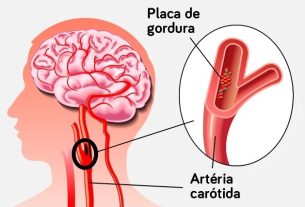
- Bone metastases, caused by various types of cancer, such as breast, prostate or lung cancer, for example;
- Changes in bone metabolism;
- Bone pain in which the cause has not been identified with other tests;
- Bone infection, such as osteomyelitis;
- Osteonecrosis;
- Arthritis;
- Primary bone tumor;
- Stress fracture or hidden fracture;
- Fractures caused by osteoporosis;
- Paget’s disease;
- Reflex sympathetic dystrophy;
- Bone infarction;
- Feasibility of bone graft.
Furthermore, scintigraphy may be recommended by the orthopedist to evaluate infections or loosening of bone prostheses.
In addition to bone scintigraphy, there are other types of scintigraphy performed on different organs of the body, such as lungs, for example, to identify various diseases. Check out how lung scintigraphy is performed.
Make an appointment with the orthopedist in the region closest to you:
Taking care of your health has never been easier!
How is the preparation
The bone scintigraphy exam generally does not require special care or fasting, and it is not necessary to suspend the use of medications that are usually used, but the doctor who will perform the exam should be informed of all the medications that are used.
To carry out the exam, it is recommended to wear comfortable clothing, without buttons or metal parts, such as zippers or belts with buckles, and you should not wear earrings, necklaces, watches or bracelets.
Furthermore, in the case of breastfeeding women, breastfeeding should be stopped on the day of the exam and for 48 hours after the exam, and avoid contact with the baby during this period.
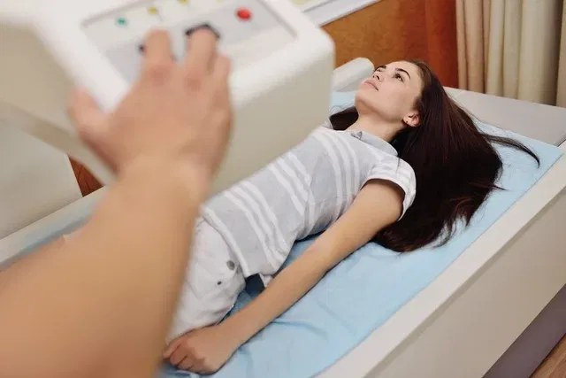
How it is made
Bone scintigraphy is carried out in the hospital or in imaging clinics, and can be carried out for a specific region or for the entire body and begins with the application of contrast containing the radiopharmaceutical, which despite being radioactive, is carried out in a safe dose for use in people.
The way the scintigraphy is performed varies depending on the type of procedure requested by the doctor, which may be:
- Conventional bone scintigraphy: After injecting the radiopharmaceutical into the vein, the person must wait around 2 to 4 hours for the radiopharmaceutical to spread throughout the body, before the image can be obtained using the scintigraphy device;
- Three-phase bone scintigraphy or bone scintigraphy with blood flow: Shortly after injecting the radiopharmaceutical, the first image is taken on the scintigraphy machine, to check the blood flow in the bones. Then, you must wait around 4 hours to obtain the second image on the scintigraphy device to check the blood balance in the bone structure and, finally, the images of radiopharmaceutical uptake by the bones are evaluated.
Between applying the radiopharmaceutical and obtaining the image on the scintigraphy machine, you must drink at least 6 to 8 glasses of water, so that the body eliminates the radiopharmaceutical that has not attached itself to the bones. In addition, you must urinate to empty your bladder before starting to obtain images on the scintigraphy equipment.
Bone scintigraphy is done in a special camera that records images of the skeleton on the computer, and the exam time is generally around 30 to 40 minutes.
In the first 24 hours after the exam, you must maintain your body’s hydration, drinking at least 8 glasses of water during this period. Furthermore, you should not come into contact with pregnant women or babies for 48 hours after the exam, as they may be sensitive to the radiopharmaceutical that is eliminated during this period.
How to understand the result
The result of the bone scintigraphy is provided by the doctor and the report describes what was observed and the images that were captured during the examination. In general, the doctor analyzes the following areas:
- Hot areaswhich are those with the most evident color, indicating that a certain region of the bone absorbed more radiation, which suggests changes, such as inflammation, fracture or bone metastases, for example.
- Cold areaswhich are those that appear clearer in the images, and indicate that there was less absorption of the radiopharmaceutical by the bones, which could mean reduced blood flow in the area or the presence of a benign tumor, for example.
The results of bone scintigraphy should always be analyzed by the doctor who requested the exam, so that the diagnosis can be made and, if necessary, the most appropriate treatment for each situation can be indicated, on an individual basis.
Who shouldn’t do
Bone scintigraphy should not be performed by women who are pregnant or suspected of being pregnant. Furthermore, you should avoid taking the exam while breastfeeding, unless necessary, and follow all medical instructions.
Bibliography
- SHIN, S. H.; KIM, S. J. Bone scintigraphy in patients with pain. Korean J Pain. 30. 3; 165-175, 2017
- VAN DEN WYNGAERT, T.; et al. EANM Bone & Joint Committee and the Oncology Committee. The EANM practice guidelines for bone scintigraphy. Eur J Nucl Med Mol Imaging. 43. 9; 1723-38, 2016
- KRUMME, JW; et al. Bone Scintigraphy: A Review of Technical Aspects and Applications in Orthopedic Surgery. Orthopedics. 42. 1; e14-e24, 2019

Sign up for our newsletter and stay up to date with exclusive news
that can transform your routine!
Warning: Undefined array key "title" in /home/storelat/public_html/wp-content/plugins/link-whisper-premium/templates/frontend/related-posts.php on line 12
Warning: Undefined array key "title_tag" in /home/storelat/public_html/wp-content/plugins/link-whisper-premium/templates/frontend/related-posts.php on line 13