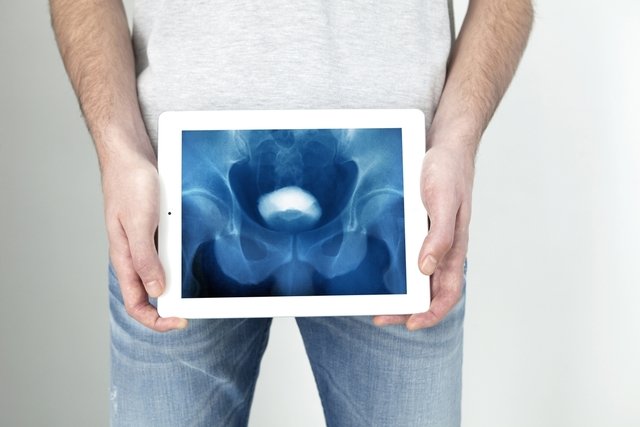
Excretory urography, also known as venous urography, is an examination performed using X-ray, computed tomography or magnetic resonance imaging, used to evaluate the structure and functions of the urinary system, including the kidneys, urethra and bladder.
This exam is indicated to identify changes such as tumors, prostate enlargement, kidney stones or hydronephrosis, an enlargement of the kidney that occurs when urine cannot reach the bladder and accumulates in the kidneys, causing pain, fever and nausea. Understand better how hydronephrosis happens.
Excretory urography lasts between 15 minutes and 4 hours and is generally carried out by a radiologist, who is responsible for carrying out tests such as computed tomography, magnetic resonance imaging and mammography, for example.

When is indicated
Excretory urography examination is generally indicated to identify changes in the urinary tract, such as the bladder, kidneys, urethra and ureter. Therefore, this examination serves to identify problems, such as:
- Tumors in the kidneys, bladder or ureter;
- Congenital diseases in the urinary system;
- Prostate enlargement;
- Hydronephrosis;
- Scars caused by infections in the urinary system;
- Kidney or bladder stones.
Excretory urography may be recommended by the doctor especially in the presence of symptoms such as pain in the lower back, presence of blood in the urine and frequent urinary infections, for example.
How is the preparation for excretory urography
Preparation for excretory urography varies according to each institution, the purpose of the examination and the age of each person. Therefore, preparation for this exam includes:
- X-ray: For this type of exam, it may be recommended to stop eating and drinking after midnight the day before the exam. In addition, the doctor may also recommend the use of a laxative medicine the night before the day of the exam;
- Computed tomography: the doctor may recommend a complete fast from food and drink for a few hours before the exam;
- Magnetic resonance imaging: It may be recommended to drink water before the exam and not urinate until the end of the exam. In addition, the doctor may also recommend fasting for 8 to 12 hours before the exam;
Furthermore, people who cannot sit still for long periods, such as children, people with claustrophobia, dementia and schizophrenia, may need sedatives to sleep during the exam. In these cases, it is important to talk to your doctor about necessary food and drink restrictions before sedation.
How the exam is carried out
Excretory urography varies according to the type of examination recommended by the doctor, and may include:
1. X-ray
During the X-ray excretory urography examination, the person lies face up, and images of the belly are taken. Next, contrast, a substance that helps to obtain better definition of images, is injected into a vein in the person’s arm or hand and other images are taken. During the exam, the radiologist may ask the person to empty their bladder to also capture an image with an empty bladder.
This exam lasts from 1 to 4 hours, and it is important to stay still while capturing the images. Additionally, the radiologist may also ask you to hold your breath for a few seconds while capturing the images.
2. MRI
During MRI, the person lies inside the device that emits the magnetic field in a prone, belly-up or side position. It is important to stay still during this exam and, therefore, the use of sedatives to sleep may be recommended for some people.
Next, the nurse or radiologist injects the contrast into the person’s vein and positions it inside the MRI machine, where the images will be taken. This exam takes approximately 15 to 30 minutes.
3. Computed tomography
In this exam, the person may lie on the table face up, face down or on their side. Next, the nurse or radiologist injects the contrast into a vein in your hand or arm. Then, the stretcher is moved into the tomograph where the images are generated.
The radiologist may also ask you to hold your breath for a few seconds during the exam. This exam lasts approximately 30 minutes. Understand better how computed tomography is performed.
During this exam it is important to remain still and, therefore, the use of sedatives to sleep may be recommended for some people.
Possible side effects
During magnetic resonance excretory urography, the part of the body being evaluated may feel slightly warm.
The contrast injection may cause a slight sting, irritation or redness at the injection site. The contrast can also cause mild symptoms that usually disappear after a few minutes, such as hot flashes, itching, nausea, headache and a metallic taste in the mouth.
Furthermore, the contrast can also cause more serious allergic reactions in some people, causing shortness of breath, vomiting, swelling in the mouth and other parts of the body, and it is recommended to immediately report these symptoms to the radiologist or nurse.
Excretory urography is not recommended for people with contrast allergies.
Risks of excretory urography
The risks of excretory urography are mainly related to the contrast injection, which can cause serious allergic reactions in some people and which includes symptoms such as swelling of the lips and throat, increased heart rate and a drop in blood pressure.
Therefore, it is recommended to be aware of possible allergy symptoms and inform the radiologist or nurse immediately. Know all the risks of using contrast.

Sign up for our newsletter and stay up to date with exclusive news
that can transform your routine!
Warning: Undefined array key "title" in /home/storelat/public_html/wp-content/plugins/link-whisper-premium/templates/frontend/related-posts.php on line 12
Warning: Undefined array key "title_tag" in /home/storelat/public_html/wp-content/plugins/link-whisper-premium/templates/frontend/related-posts.php on line 13


