The proctological examination is a type of examination that involves external evaluation of the region, rectal examination, anoscopy and rectosigmoidoscopy. This exam is carried out with the aim of investigating gastrointestinal changes and identifying fissures, fistulas and hemorrhoids, in addition to being an important exam used to prevent colorectal cancer.
The proctological examination is carried out in the office and lasts around 10 minutes, with no preparation necessary for it. Despite being simple, it can be uncomfortable, especially if the person has anal fissures or hemorrhoids.
This exam is indicated for all people who are suspected of having changes in the anus or rectum, and it is important that it is carried out so that the diagnosis can be made and the most appropriate treatment can be initiated.

What is it for
Proctological examination is normally carried out with the aim of:
- Prevent colorectal cancer;
- Diagnose internal and external hemorrhoids;
- Investigate the presence of anal fissures and fistulas;
- Identify the cause of anal itching;
- Check for the presence of anorectal warts;
- Investigate the cause of blood and mucus in the stool.
It is important that the proctological examination is carried out as soon as the person identifies any anorectal signs or symptoms, such as anal pain, presence of blood and mucus in the stool, pain and difficulty in evacuating and anal discomfort.
The proctological examination is carried out by a proctologist or gastroenterologist to identify changes in the anal and rectal canal that can be very uncomfortable and have a negative impact on the person’s life.
How is done
Before starting the exam itself, an assessment of the signs and symptoms described by the person is carried out, in addition to evaluating the clinical history, lifestyle habits and intestinal routine, so that the doctor can conduct the exam in the best way.
The proctological examination is carried out in stages, and it is initially recommended that the person put on an appropriate gown and lie on their side with their legs curled up. The doctor then begins the examination, which, in general, can be divided into external evaluation, rectal examination, anoscopy and rectosigmoidoscopy:
1. External evaluation
External evaluation is the first stage of the proctological examination and consists of the doctor observing the anus with the aim of checking for the presence of external hemorrhoids, fissures, fistulas and dermatological changes that cause anal itching. During the evaluation, the doctor may also ask the person to strain as if they were going to have a bowel movement, as this way it is possible to check if there are swollen veins that are indicative of grade 2, 3 or 4 internal hemorrhoids.
2. Digital rectal examination
In this second stage of the examination, the doctor performs a rectal examination, in which the index finger is inserted into the person’s anus, duly protected by a glove and lubricated, with the aim of evaluating the anal orifice, the sphincters and the final part of the intestine, making it possible to identify the presence of nodules, fistulous orifices, feces and internal hemorrhoids.
Furthermore, through a rectal exam, the doctor can check for the presence of palpable anal lesions and the presence of blood in the rectum. Understand how a rectal examination is performed.
3. Anuscopy
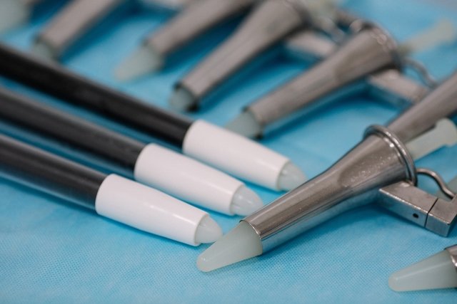
Anoscopy allows you to better visualize the anal canal, making it possible to identify changes that were not detected by rectal examination. In this examination, a medical device called an anoscope is introduced into the anus, which is a disposable transparent or metal tube that must be properly lubricated before it can be inserted into the anus.
After insertion into the anoscope, light is applied directly to the anus so that the doctor can better visualize the anal canal, making it possible to identify hemorrhoids, anal fissures, ulcers, warts and signs indicative of cancer.
4. Retossigmoidoscopia
Rectosigmoidoscopy is only indicated when other tests have not been able to identify the cause of the signs and symptoms presented by the person. Through this examination, it is possible to visualize the final portion of the large intestine, identifying changes and signs indicative of disease.
In this exam, a rigid or flexible tube is introduced into the anal canal with a microcamera at its end, making it possible for the doctor to make a more precise assessment of the region and be able to more easily identify changes such as polyps, lesions, tumors or foci of bleeding. See how rectosigmoidoscopy is performed.
Bibliography
- GOVERNMENT OF THE STATE OF ESPÍRITO SANTO. Regulatory protocols for access to specialized proctology consultations and exams. 2017. Available at: <https://saude.es.gov.br/Media/sesa/Protocolo/Protocolo%20Proctologia.pdf>. Accessed on January 7, 2020

Sign up for our newsletter and stay up to date with exclusive news
that can transform your routine!
Warning: Undefined array key "title" in /home/storelat/public_html/wp-content/plugins/link-whisper-premium/templates/frontend/related-posts.php on line 12
Warning: Undefined array key "title_tag" in /home/storelat/public_html/wp-content/plugins/link-whisper-premium/templates/frontend/related-posts.php on line 13


