Echo Doppler is a type of ultrasound, with specific techniques, that allows color visualization of the blood flow in the body’s arteries and veins, helping to check the functioning of tissues, such as the walls of the heart, nerves and brain.
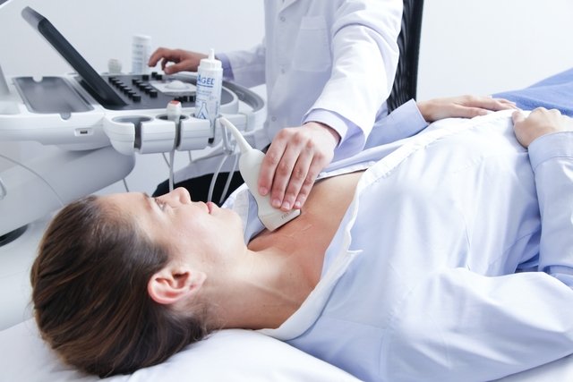
It is a type of non-invasive exam, that is, it does not use needles and does not require anesthesia to be carried out, and is carried out by a radiologist, who will pass a transducer with gel, which is a small part of the ultrasound device, into the location of the body to be examined.
Using Doppler ultrasound can diagnose various diseases such as atherosclerosis, vasculitis and aneurysms, which is why it is often recommended by a cardiologist or neurologist. However, this exam is also recommended by obstetricians in order to check the baby’s health conditions during pregnancy.

What is it for
Echo Doppler is a type of ultrasound that is used to check blood flow in the veins and arteries, heart, brain and even the lower limbs. Therefore, this exam may be indicated for the following situations:
- Detect obstructions due to fat in arteries or veins;
- Locate blood clots in the veins of the arm or leg;
- Check whether there is any dilation of the walls of the veins or arteries;
- Analyze the results of heart surgeries;
- Assess the characteristics of varicose veins.
Furthermore, the Doppler ultrasound can also help check blood pressure within the arteries, showing the amount of blood flowing in the blood vessels and can be done as an alternative to other more invasive tests, such as angiography, which involves injecting contrast into the vein.
This exam can also be performed in children and is generally recommended by the pediatrician to assess whether there is any malformation in the heart or to assist in the placement of a central venous catheter. See more about what a central venous catheter is and in which cases it is indicated.
How is done
The Doppler ultrasound exam is carried out by a radiologist in a room at a unit, or diagnostic center, and does not require anesthesia or intravenous contrast, in addition to not using any type of radiation.
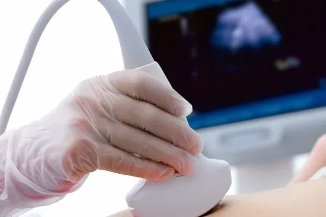
To take the exam, the person must put on an apron and lie on a stretcher. Then the doctor will apply a gel and move a transducer across the skin, which is a small device with which it will be possible to visualize the internal parts of the body, such as veins and arteries. This causes no pain or discomfort.
The doctor will view the images on a computer screen and analyze the body’s structures, and after a few days, a report will be issued with a description of what was found in the examination and this report must be delivered to the doctor who requested it.
Preparation for the exam
In most cases, no specific care is needed to perform the exam, however, people who use medications that alter blood pressure or who smoke should inform the doctor who will perform the exam, as these situations can increase blood flow. of blood in the body’s veins and arteries.
What are the types of echo Doppler
Depending on the part or structure of the body that the doctor wants to be analyzed, the exam may be:
- Ecodoppler fetal: carried out during pregnancy, it consists of a cardiac assessment of the baby;
- Lower limb Doppler ultrasound: serves to analyze veins and arteries in the legs;
- Upper limb Doppler ultrasound: consists of checking the condition of the veins and arteries in the arms;
- Carotid echodoppler: indicated to check the vein that supplies blood to the head region;
- Doppler ultrasound of renal arteries: recommended for analysis of kidney veins and arteries;
- Ecodoppler transcraniano: recommended to evaluate the veins and arteries of the brain;
- Thyroid Doppler: It is the type that is used to check blood flow in the thyroid.
These specific types of Doppler ultrasound can be requested when consulting a cardiovascular doctor or neurologist, but they can also be indicated for people who are admitted to a hospital with suspected disease or alteration.
Main diseases diagnosed
Echo Doppler, or Doppler ultrasound, may be recommended by a cardiovascular doctor, neurologist or nephrologist to investigate and diagnose some diseases such as:
1. Atherosclerosis
Atherosclerosis is a disease that occurs due to the accumulation of fatty plaques, or atheromas, in the arteries of the heart and which, if left untreated, can block blood flow and lead to serious complications such as acute myocardial infarction and accidents. cerebrovascular.
Doppler echocardiography is a type of examination widely used to investigate this disease, however, the cardiologist may request other examinations such as angiography and cardiac catheterization. After diagnosing this change, the doctor will recommend the most appropriate treatment, which is based on changes in habits and medications. See other treatment options for atherosclerosis.
2. Vasculitis
Vasculitis is a change caused by inflammation of the body’s blood vessels and can cause symptoms such as red spots on the skin, tingling or loss of sensation in the hands or feet, joint pain and fever. This disease can be caused by other conditions such as infections, autoimmune diseases and cancer and, in some cases, lead to complications such as bleeding.
A rheumatologist should be consulted if vasculitis is suspected, and he or she may recommend a Doppler ultrasound to confirm the diagnosis. Treatment of this disease is recommended by the doctor according to the severity and location of the inflammation of the blood vessels. Check out other tests that can be done to confirm the diagnosis of vasculitis and treatment.
3. Aneurysms
Aneurysms can arise due to an increase in the pressure at which blood passes through a blood vessel, which leads to the formation of a dilation of the wall of the vein or artery. This dilation can occur in blood vessels in the heart, brain or in parts of the body, such as the abdominal aorta, for example.
Symptoms depend on the location of the aneurysm, and people who suffer this change may have intense pain in the area, difficulty walking, tingling in the head, blurred vision and even seizures and must seek emergency care at a hospital. Check out the main symptoms of cerebral and aortic aneurysm.
4. Deep vein thrombosis
Deep vein thrombosis is a situation that occurs due to an obstruction of a deep vein in the leg, thigh or abdomen, compromising blood flow and, in most cases, causing swelling, intense pain and a purple color in the leg, for example.
Some risk factors are related to the appearance of deep vein thrombosis, such as cancer, major surgery, use of oral contraceptives and little movement of the body, and the diagnosis is made through Doppler ultrasound. Hospitalization is often necessary for the treatment of this change, which is based on the use of anticoagulant medications, such as heparin. Find out how to avoid thrombosis in the leg.
5. Renal artery stenosis
Renal artery stenosis is defined as the narrowing of the main artery of the kidneys due to fatty plaques, blood clots or tumors and the diagnosis of this change is made through tests such as angiography and renal Doppler ultrasound.
The treatment of renal artery stenosis is recommended by a nephrologist and consists of catheterization, surgery and the use of anticoagulant and thrombolytic medications. Often, this treatment must be carried out with the person admitted to a hospital so that they can receive the medication via vein and must be started as soon as possible to avoid complications such as pulmonary edema.

Sign up for our newsletter and stay up to date with exclusive news
that can transform your routine!
Warning: Undefined array key "title" in /home/storelat/public_html/wp-content/plugins/link-whisper-premium/templates/frontend/related-posts.php on line 12
Warning: Undefined array key "title_tag" in /home/storelat/public_html/wp-content/plugins/link-whisper-premium/templates/frontend/related-posts.php on line 13