Pyelocaliceal dilation, also known as renal calyceal ectasia, hydronephrosis or dilated kidney, is a dilation or enlargement of the internal portion of the kidney. This region is known as the renal pelvis, as it is shaped like a funnel and has the function of collecting urine and taking it towards the ureters and bladder.
Generally, pyelocaliceal dilation occurs due to a blockage in the passage of urine, which leads to retention of urine in the renal pelvis and kidneys, which can be caused by deformities in the structures of the urinary tract in babies or even by stones, cysts, tumors or infection. serious damage to the kidneys, which can also occur in adults.
Pyelocaliceal dilation does not always cause symptoms, but pain in the abdomen, difficulty urinating or blood in the urine may appear, for example, and it is important to consult a pediatrician, general practitioner or nephrologist to identify the cause and provide the most appropriate treatment.

Main symptoms
The main symptoms of pyelocaliceal dilation include:
- Severe and sudden pain in the lower back, which may worsen after drinking liquid;
- Constant pain in the upper abdomen and back;
- Nausea and vomiting;
- Need to urinate more frequently;
- Pain or burning when urinating;
- Feeling of a full bladder even after urinating;
- Difficulty urinating;
- Reduction in urine volume;
- Blood in the urine;
- Low fever;
- Bad to be.
Symptoms may vary according to the cause of the dilation and signs or symptoms are not always noticed.
In addition, pyelocaliceal dilation in babies may also have no symptoms, however, a pediatrician should be seen as soon as possible if the baby has a high fever for no apparent reason, crying when urinating or urine that is darker than normal, as this can indicate urinary infection and must be treated immediately. See other symptoms of urinary tract infection in babies.
How to confirm the diagnosis
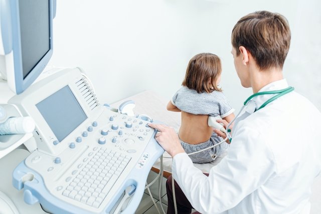
The diagnosis of pyelocaliceal dilation is made by the doctor through an ultrasound or ultrasound examination of the renal system, which can demonstrate the degree of dilation, the size of the kidney and whether its size causes compression of the kidney tissues.
In some cases, dilation can be detected in the baby while still in the womb, during routine ultrasound exams, but it is normally confirmed after the baby is born.
Other tests that may be recommended by the doctor are excretory urography, voiding urethrography or renal scintigraphy, for example, which can evaluate more details of the anatomy and flow of urine through the urinary tract. Understand how it is performed and the indications for excretory urography.
Make an appointment with your nearest doctor to further evaluate your kidneys:
Taking care of your health has never been easier!
Possible causes
Pyelocaliceal dilation occurs due to an obstruction of the passage of urine through the pyelocaliceal system and can occur in newborns or adults, the main causes of which include:
- Pyelocaliceal dilation in the newborn: Its causes are not yet fully understood and, in most cases, it tends to disappear after the birth of the baby. However, there are cases caused by anatomical deformities in the urinary tract, which are more serious situations and can cause recurrent urinary infections;
- Pyelocaliceal dilation in adults: It generally occurs as a result of cysts, stones, nodules or cancer in the region of the kidneys or ureters, which block the passage of urine and accumulate it, causing dilation of the renal pelvis. Check out more causes of pyelocaliceal dilation in adults.
Pyelocaliceal dilation generally occurs more frequently in the right kidney, but it can also occur in the left kidney, or in both kidneys, being bilateral.
Is pyelocaliceal dilation serious?
If left untreated, pyelocaliceal dilation can be serious, as it can cause kidney failure and alter or permanently damage the functioning of the kidneys.
How the treatment is carried out
Treatment for pyelocaliceal dilation in newborns depends on the size of the dilation. When the dilation is less than 10 mm, the baby only needs to have several ultrasounds for the pediatrician to monitor its progress, as, normally, the dilation tends to disappear.
When dilation is greater than 10 mm, treatment is with antibiotics prescribed by the pediatrician. In more serious cases, where dilation is greater than 15 mm, surgery is recommended to correct the cause of dilation.
In adults, treatment for pyelocaliceal dilation can be done with medications prescribed by a urologist or nephrologist, and surgery may be necessary, depending on the kidney disease that caused the dilation.

Bibliography
- SZKODZIAK, P. Ultrasound screening for pyelectasis in pregnant women. Clinical necessity or “art for art’s sake”?. J Ultrasound. 18.73; 152-157, 2018
- DRNASIN, K.; et al. Clinical importance of pyelocalyceal dilation diagnosed by postnatal ultrasonographic screening of the urinary tract. Med Sci Monit. 19. 125–131, 2013

Sign up for our newsletter and stay up to date with exclusive news
that can transform your routine!
Warning: Undefined array key "title" in /home/storelat/public_html/wp-content/plugins/link-whisper-premium/templates/frontend/related-posts.php on line 12
Warning: Undefined array key "title_tag" in /home/storelat/public_html/wp-content/plugins/link-whisper-premium/templates/frontend/related-posts.php on line 13